Trochanteric Fracture in Hauts-de-Seine

Hip Surgery

What Is a Trochanteric Fracture?

Dr Simon Tournemine in Hauts-de-Seine (92)

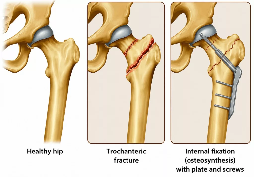
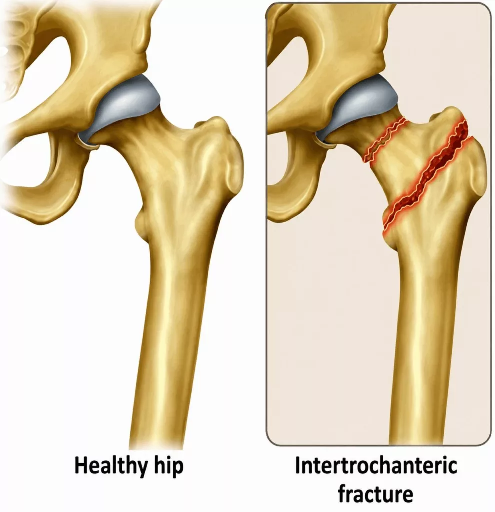
A trochanteric fracture is a break in the continuity of the bone involving the trochanteric region of the femur. This area, located between the femoral shaft and the femoral neck, is particularly vulnerable to fracture. Once fractured, the femur can no longer support the body’s weight, making standing and walking impossible while causing severe pain around the hip.

Causes and Risk Factors for a Trochanteric Fracture

Hip Surgery Specialist in Hauts-de-Seine (92)

More than 60,000 trochanteric fractures occur each year in France. Although younger individuals may sustain this type of injury, the vast majority of patients are over the age of 60.

Women are more frequently affected than men, particularly after the menopause, when osteoporosis, a condition that weakens bone strength, becomes more common.

In younger patients, trochanteric fractures usually result from high-energy trauma, most commonly road traffic accidents. In older adults, however, a simple fall may be sufficient to cause a fracture of the trochanteric region.

Certain conditions, including osteoporosis and other diseases associated with reduced bone strength, increase the risk of fracture following a fall.

Trochanteric Fracture in Rueil-Malmaison

Hip Surgery in Hauts-de-Seine

A trochanteric fracture most commonly occurs following a fall. Severe pain develops immediately around the hip, making it impossible to stand or move the affected leg. A visible deformity may be present, with the injured limb appearing shortened and malpositioned. Bruising around the hip may also develop.

In rare cases, when the fracture is stable and undisplaced, there may be no visible deformity and the patient may still be able to walk despite the pain.

Although not all trochanteric fractures can be prevented, reducing recognised risk factors can help lower the likelihood of injury. Preventive measures include:

  • screening for and treating osteoporosis;
  • achieving a healthy weight in cases of overweight or obesity;
  • maintaining a healthy, balanced diet and engaging in regular physical activity to preserve bone strength and overall physical condition;
  • identifying and managing factors that increase the risk of falls, including visual, hearing and balance disorders;
  • avoiding smoking and excessive alcohol consumption.

The consultation begins with a detailed medical history to establish the circumstances surrounding the fall or injury.

Clinical examination typically reveals severe hip pain associated with an inability to stand or move the affected leg. A deformity of the injured limb may be visible, with the leg appearing shortened and malpositioned. Bruising may also be present.

In rare cases, when the fracture is stable and undisplaced, no obvious deformity is visible and walking may still be possible despite the pain.

A thorough neurological and vascular examination is performed to identify any associated injury to nearby nerves or blood vessels. The assessment also looks for the presence of other fractures that may have occurred at the same time.

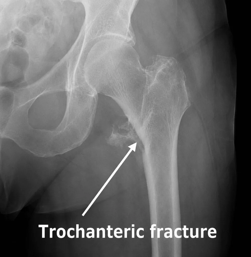
Standard pelvic X-rays confirm the diagnosis by demonstrating a break in the continuity of the trochanteric region, often associated with displacement of the trochanter and femoral neck. Additional imaging is not usually required.
Without surgical treatment, the natural course of a trochanteric fracture is generally unfavourable, with persistent pain and an inability to resume walking.

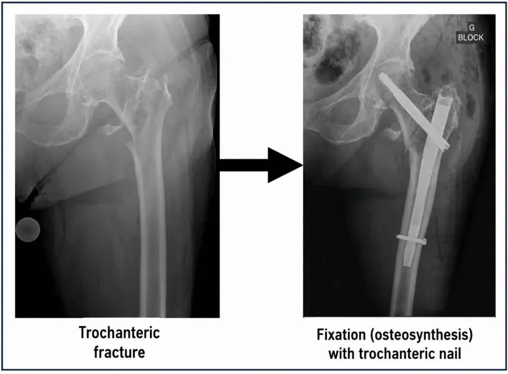
The standard treatment is internal fixation (osteosynthesis). During the procedure, the fractured bone fragments are repositioned and stabilised using either a cephalomedullary (trochanteric) nail or a sliding hip screw and plate. Both techniques are designed to stabilise the fracture, promote bone healing and allow early mobilisation. The choice between these surgical options depends on the fracture pattern and should be discussed with your orthopaedic surgeon.