
Patellar Instability in Hauts-de-Seine

Knee Surgery

What Is Patellar Instability?

Dr Simon Tournemine in Hauts-de-Seine (92)

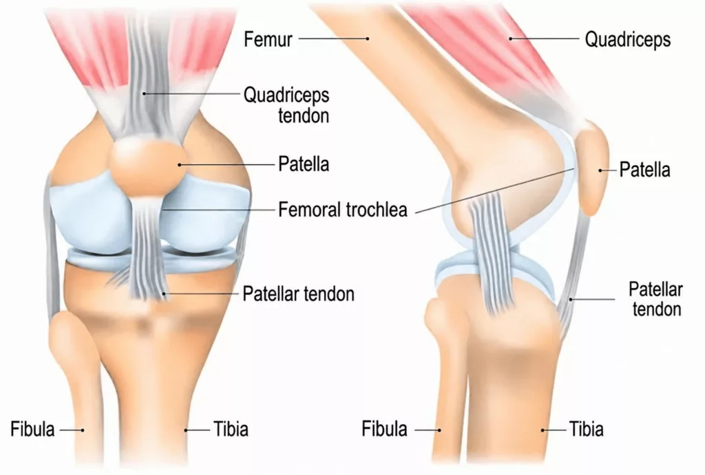
The patella (kneecap) is the third bone of the knee joint, alongside the femur and the tibia. Located at the front of the knee, it is embedded within the extensor mechanism, between the quadriceps tendon above and the patellar tendon below. When the quadriceps muscle contracts, it pulls on the quadriceps tendon, the patella and the patellar tendon, allowing the knee to straighten.

During knee flexion, the patella glides along the front of the femur within a groove known as the femoral trochlea, which guides its movement throughout the range of motion. Patellar instability occurs when the patella no longer tracks correctly within this groove, creating the sensation that the kneecap is slipping or giving way. In most cases, the patella has a tendency to shift towards the outside of the knee (lateral instability).

Causes and Risk Factors for Patellar Instability

Knee Surgery Specialist in Hauts-de-Seine (92)

Patellar dislocation is a common reason for orthopaedic consultation, particularly among young women and physically active individuals.

There are two principal causes of patellar instability: post-traumatic instability and developmental instability.

Post-traumatic patellar instability develops following a knee injury that forces the patella outwards (lateral dislocation), resulting in tearing of the medial patellofemoral ligament (MPFL). Once this ligament has been injured, the patella is no longer adequately restrained, making recurrent instability more likely.

Developmental patellar instability is related to anatomical features that predispose the patella to recurrent dislocation. These include:

  • Trochlear dysplasia: the femoral trochlea, which normally guides the patella during knee movement, is too shallow and therefore provides insufficient stability.
  • Patella alta: the patella sits higher than normal because the patellar tendon is excessively long, increasing the risk of abnormal tracking and dislocation.
  • Excessive lateral position of the tibial tubercle: because the patellar tendon inserts onto the tibial tubercle, an excessively lateral position alters the alignment of the extensor mechanism and pulls the patella outwards during knee flexion.

Patellar Instability in Rueil-Malmaison

Knee Surgery in Hauts-de-Seine

Each episode of patellar dislocation causes the kneecap to move out of its normal groove, resulting in sudden, intense pain, swelling of the knee and, in some cases, a fall. Sporting activity cannot usually be continued after the injury.

Over time, episodes of instability often become more frequent and may occur after increasingly minor trauma, causing significant limitations in everyday life. Participation in sport is frequently affected. Many patients also experience apprehension, a sensation that the patella is shifting outwards without fully dislocating, giving the impression that the knee cannot be controlled properly.

With each episode of instability, the abnormal movement of the patella against the femur progressively damages the cartilage of both the patella and the femoral trochlea. Over time, this may lead to patellofemoral osteoarthritis, causing chronic pain at the front of the knee. These symptoms are typically aggravated by climbing or descending stairs, prolonged sitting or walking on sloping ground.

The consultation begins with a detailed medical history to determine how the instability developed, the number of previous dislocations and the circumstances in which they occurred. Apprehension is one of the most common symptoms reported by patients and often leads to reduced participation in sporting activities.

Clinical examination looks for anatomical factors associated with patellar instability, including generalised ligamentous laxity, excessive patellar mobility and genu valgum (knock knees). The Smillie apprehension test is particularly sensitive. During this manoeuvre, the patella is gently displaced laterally while the knee is flexed, reproducing the sensation experienced during episodes of instability.

Imaging studies are systematically performed to identify anatomical risk factors and assess any bone or ligament injuries resulting from previous dislocations.

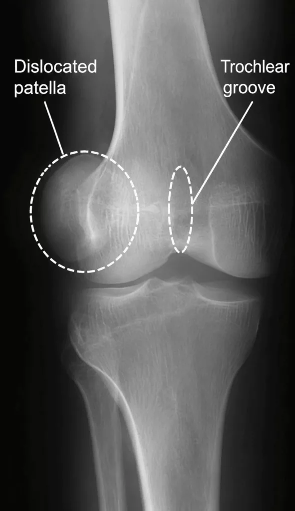
Standard knee X-rays are used to measure patellar height, evaluate trochlear dysplasia and identify any associated fractures.

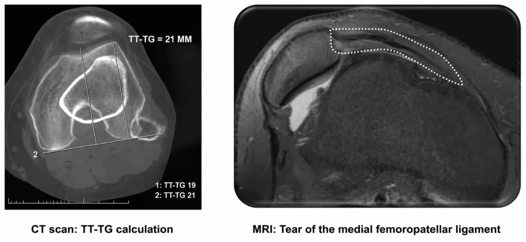
A CT scan is essential for measuring the tibial tubercle-trochlear groove (TT-TG) distance, assessing trochlear dysplasia and evaluating patellar tilt.

Magnetic resonance imaging (MRI) may also be indicated to assess the integrity of the medial patellofemoral ligament (MPFL) and evaluate cartilage damage resulting from recurrent episodes of patellar dislocation.
Surgical treatment for patellar instability is generally reserved for patients who have experienced at least one episode of patellar dislocation and/or who present with anatomical abnormalities that predispose them to recurrent instability.

When these criteria are not present, conservative management is recommended. This consists of a structured physiotherapy programme focusing on strengthening the muscles around the knee, improving patellar tracking and reducing symptoms.