High Tibial Valgus Osteotomy in Hauts-de-Seine
Knee Surgery
What Is a High Tibial Valgus Osteotomy?
Dr Simon Tournemine in Hauts-de-Seine (92)
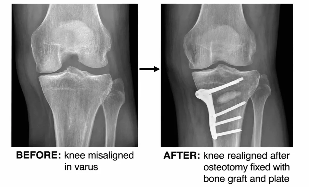
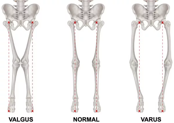
A high tibial valgus osteotomy (HTO) is a joint-preserving surgical procedure that involves cutting the upper part of the tibia to correct abnormal lower limb alignment. When the legs are aligned in varus (bow-legged) or valgus (knock-kneed), the mechanical load is distributed unevenly across the knee joint, leading to pain and accelerated cartilage wear (osteoarthritis).
By restoring the normal alignment of the lower limb, weight-bearing forces are redistributed more evenly across the knee. This helps relieve pain, slows the progression of osteoarthritis and preserves the native joint for longer. This procedure is particularly suitable for patients under the age of 60 with early to moderate osteoarthritis. After the age of 65, a unicompartmental knee replacement (UKR) is generally considered instead.
High Tibial Valgus Osteotomy Procedure
Knee Surgery Specialist in Hauts-de-Seine (92)
This procedure requires highly accurate correction of the limb alignment. A three-dimensional CT-based preoperative plan is therefore performed, and patient-specific cutting guides are manufactured to match your anatomy precisely and achieve the planned correction.
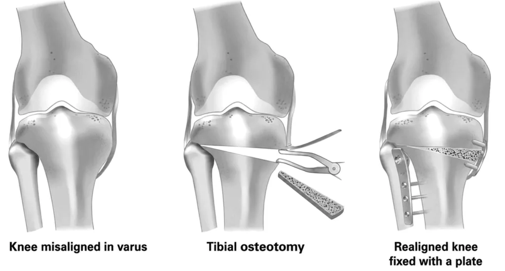
An incision of approximately 10 cm is made over the upper part of the tibia. The patient-specific guides are positioned to perform the planned bone cut. The osteotomy is then gradually opened to restore the desired alignment of the leg according to the preoperative plan.
The corrected position is stabilised using a metal plate and high-strength screws. The opening created during the osteotomy is filled with a bone substitute graft to promote bone healing.
The procedure usually lasts approximately 40 minutes.
It is performed under either general anaesthesia or spinal anaesthesia, during which only the lower limbs are anaesthetised. The type of anaesthesia is chosen jointly with your anaesthetist during the preoperative consultation.
This operation may be performed as day-case surgery, allowing discharge on the same day, or may require one overnight stay, depending on the individual case.
High Tibial Valgus Osteotomy in Rueil-Malmaison
Knee Surgery in Hauts-de-Seine
When Is a High Tibial Valgus Osteotomy Recommended?
Knee osteoarthritis (gonarthrosis) is a progressive degenerative condition for which no treatment can restore damaged cartilage. Conservative management initially includes lifestyle modifications, pain-relieving medication, anti-inflammatory treatment and physiotherapy to reduce symptoms.
When these treatments no longer provide sufficient relief and knee pain begins to interfere with everyday activities, a high tibial valgus osteotomy may be considered.
This procedure is particularly effective for early-stage osteoarthritis in patients younger than 65 years. In more advanced osteoarthritis or in patients over the age of 65, a unicompartmental knee replacement (UKR) is generally the preferred option. Consequently, consideration of high tibial osteotomy should occur relatively early in the course of the disease.
Other factors must also be considered, including the severity of the deformity, the presence of associated ligament injuries, the patient’s activity level and their medical history.
The choice between high tibial valgus osteotomy and unicompartmental knee replacement is therefore individualised and should be discussed thoroughly with your orthopaedic surgeon.
Recovery and Rehabilitation After High Tibial Valgus Osteotomy
Following surgery, the dressing is generally changed every two days for approximately two weeks. Pain management is closely monitored to maximise comfort and facilitate early rehabilitation. Anticoagulant treatment is prescribed for six weeks to reduce the risk of deep vein thrombosis.
After the operation, the knee is supported in a hinged brace, and walking is permitted using two crutches with partial weight-bearing for six weeks.
Enhanced Recovery After Surgery (ERAS) protocols are used whenever appropriate. A physiotherapist will assist you in standing, walking and beginning rehabilitation on the day of surgery. Early mobilisation combined with physiotherapy plays a key role in preserving muscle strength and restoring knee mobility.
Following discharge, your surgeon will provide all necessary prescriptions, including dressings, pain medication, anticoagulants and physiotherapy. Rehabilitation should continue either at home with a physiotherapist or within a rehabilitation centre. Early resumption of daily activities remains an essential component of recovery.
Driving is generally possible after approximately six weeks. Sick leave is also usually around six weeks, although this varies according to the patient’s occupation, with office-based work often resumed sooner. Sporting activities are gradually reintroduced from the third postoperative month.
These recovery times are average estimates and vary between individuals.
Regular long-term clinical and radiographic follow-up is essential to monitor bone healing and assess the condition of the knee.
Risks and Complications of High Tibial Valgus Osteotomy
Every effort is made to ensure a successful recovery. Nevertheless, as with any surgical procedure, complications, although uncommon, may occur.
The principal risks include:
- Deep vein thrombosis and pulmonary embolism: uncommon complications, with the risk reduced by anticoagulant treatment prescribed after surgery;
- Exacerbation of pre-existing medical conditions, such as heart failure or diabetes. Careful postoperative monitoring by the anaesthetic team helps minimise these risks;
- Infection (risk below 1%). This may require further surgery to wash out the surgical site, followed by antibiotic treatment. As smoking significantly increases both infection rates and wound-healing complications, patients are strongly advised to stop smoking at least one month before surgery and to continue abstaining for three months afterwards;
- Reduced knee mobility and postoperative stiffness, particularly if rehabilitation is not carried out adequately;
- Haematoma, which may occasionally require drainage or blood transfusion if significant postoperative bleeding occurs;
- Sensory nerve injury, involving small cutaneous nerve branches around the knee, potentially causing localised numbness;
- Non-union (pseudarthrosis): failure of the osteotomy to heal, which may require additional surgery to stimulate bone union;
- Complex Regional Pain Syndrome (CRPS): a rare inflammatory pain condition with an unpredictable course that may require specialised rehabilitation and pain management;
- Injury to the common fibular nerve or tibial nerve: an exceptionally rare complication that may occur following a haematoma, surgical trauma or regional anaesthesia. Partial or complete recovery may occur over several months;
- Injury to the blood vessels of the leg: a very rare complication that may require urgent vascular surgery to restore blood flow;
- Compartment syndrome: increased pressure within the muscles of the leg, usually caused by bleeding, which compromises the local blood supply and requires emergency surgical decompression.
These represent the principal risks associated with high tibial valgus osteotomy, but the list is not exhaustive. Other exceptional complications may occur. Your orthopaedic surgeon will discuss your individual situation and answer any specific questions before surgery.
Results of High Tibial Valgus Osteotomy
The natural progression of osteoarthritis in a malaligned knee is unfavourable. Progressive deformity increases abnormal loading across the joint, leading to worsening cartilage damage, meniscal degeneration and osteoarthritis, which may ultimately require knee replacement surgery.
High tibial valgus osteotomy has two principal objectives:
- To restore normal limb alignment, relieving pain and feelings of instability while allowing patients to return to sporting activities;
- To redistribute mechanical forces across the knee, slowing the progression of cartilage wear and helping preserve the native joint over the long term.
Thanks to patient-specific 3D planning, highly accurate correction can be achieved, providing very good outcomes in the majority of appropriately selected patients.
Most patients progressively resume their personal, professional and sporting activities after approximately six weeks. Once rehabilitation has been completed and bone healing is achieved, all sporting activities may generally be resumed without restriction.