Patellar Stabilisation in Hauts-de-Seine
Knee Surgery
What Is Patellar Stabilisation?
Dr Simon Tournemine in Hauts-de-Seine (92)
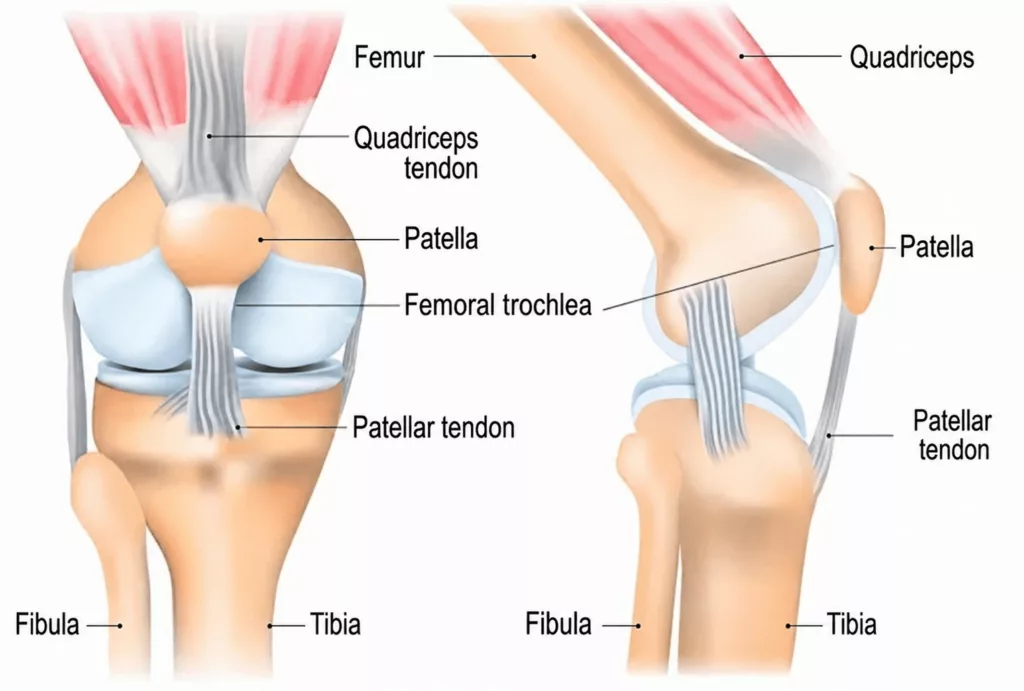
The patella, or kneecap, is the third bone of the knee joint, together with the femur and tibia. It is located at the front of the knee and is incorporated into the extensor mechanism between the quadriceps tendon and the patellar tendon.
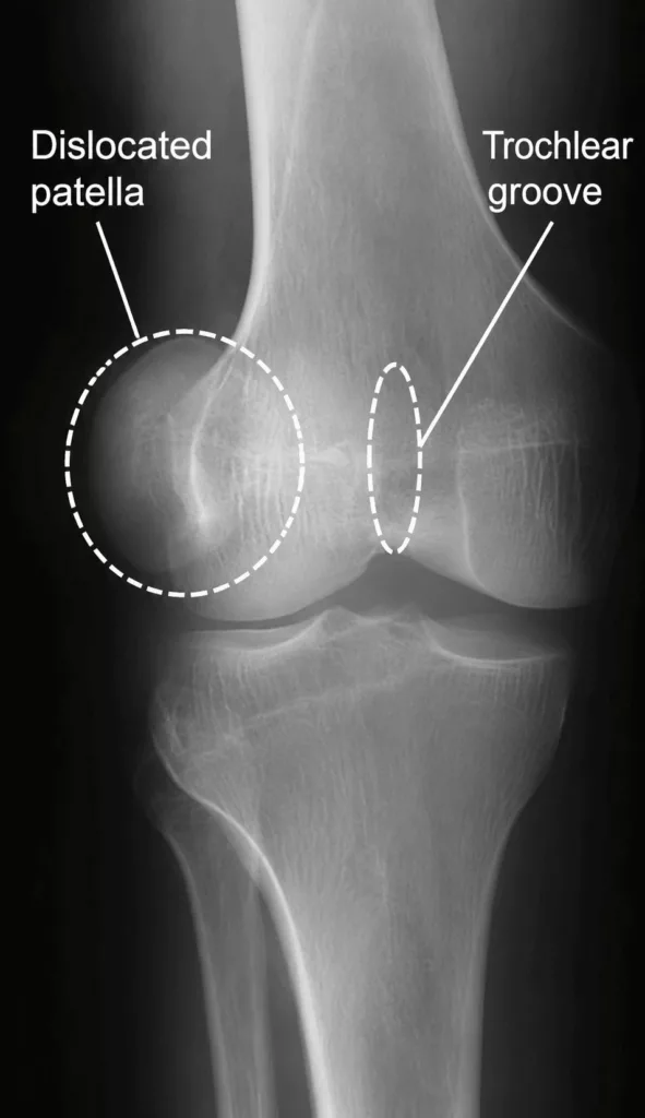
During knee flexion, the patella glides over the front of the femur and engages within a groove known as the femoral trochlea, which guides its movement. Patellar instability occurs when the kneecap does not track correctly within the trochlear groove, creating a sensation that it is slipping or moving out of place. In most cases, the patella tends to shift towards the outside of the knee.
There are two principal causes of patellar instability:
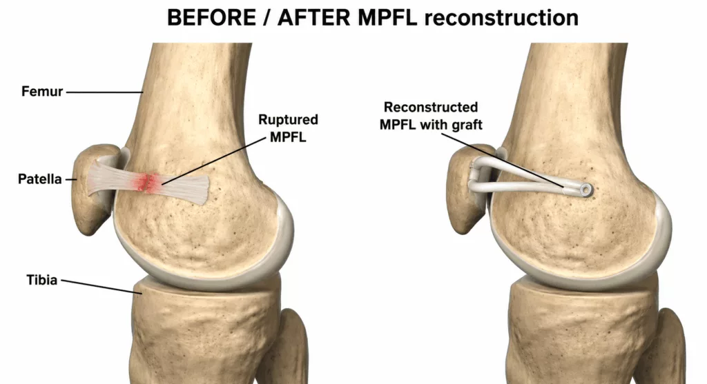
Post-traumatic instability: patellar instability develops following a knee injury that forces the patella outwards and tears the medial patellofemoral ligament (MPFL). Once this ligament has been damaged, the patella is no longer adequately restrained. Reconstruction of the medial patellofemoral ligament may then be performed to restore stability.
Developmental instability: patellar instability is related to the anatomical morphology of the knee, which predisposes the patient to dislocation.
The principal anatomical factors include:
- Trochlear dysplasia: the trochlea is the groove within which the patella glides over the femur. When this groove is too shallow, it provides insufficient guidance and increases the risk of patellar dislocation.
- Patella alta: the patella sits too high in relation to the tibia because the patellar tendon is excessively long, making abnormal tracking more likely.
- Excessive lateral position of the tibial tubercle: the patellar tendon inserts onto the tibial tubercle. When this insertion is positioned too far laterally, it pulls the patella outwards during knee flexion and may cause it to dislocate.
In these situations, a tibial tubercle osteotomy may be performed to reposition the tubercle appropriately.
Patellar Stabilisation Procedure
Knee Surgery Specialist in Hauts-de-Seine (92)
The procedure selected depends on the underlying cause of the instability. Treatment may involve medial patellofemoral ligament reconstruction, tibial tubercle osteotomy and repositioning, or a combination of both procedures.
Patellar Stabilisation in Rueil-Malmaison
Knee Surgery in Hauts-de-Seine
Patellar Stabilisation Surgery
Medial Patellofemoral Ligament Reconstruction
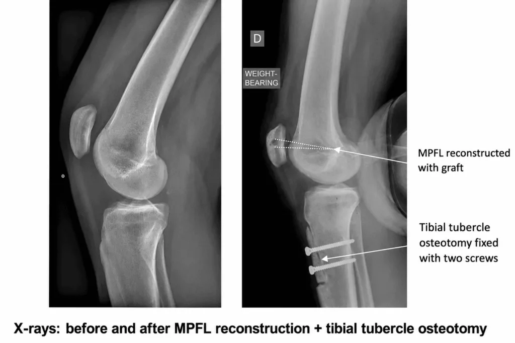
This procedure consists of replacing the torn ligament with a new ligament graft. A hamstring tendon, usually the gracilis tendon, is harvested and positioned between the femur and the patella to replace the damaged medial patellofemoral ligament. The graft is secured to the bone using two interference screws.
The lateral patellofemoral ligament is also divided to help recentre the patella.
Harvesting a hamstring tendon has minimal functional consequences, as the donor site heals progressively over the following weeks.
Three small incisions are required: one to harvest the tendon, one to secure the graft to the femur and one to fix it to the patella.

Tibial Tubercle Osteotomy and Repositioning
This procedure aims to reposition the patella in relation to the trochlear groove within which it moves.
The tibial attachment of the patellar tendon, known as the tibial tubercle, is cut, repositioned and secured to the tibia using two screws. This allows the patella to be lowered when it sits too high and moved medially when it is positioned too far laterally.
An incision of approximately 10 cm is made over the front of the tibia.
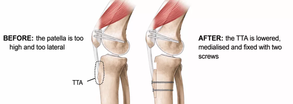
Both procedures are frequently combined.
These operations generally last approximately 40 minutes.
They are performed under either general anaesthesia or spinal anaesthesia, in which only the lower limbs are anaesthetised. The choice of anaesthetic is made jointly with the anaesthetist during the preoperative consultation.
These procedures are usually performed as day-case surgery, allowing patients to return home on the same day.
Postoperative Care and Rehabilitation After Patellar Stabilisation
The dressing is generally changed every two days for approximately two weeks. Pain management is carefully monitored to maximise comfort and facilitate early rehabilitation.
Anticoagulant treatment is prescribed for two weeks to reduce the risk of deep vein thrombosis. This medication is taken in tablet form and does not require specific monitoring.
Rehabilitation differs according to the procedure performed: tibial tubercle transposition or MPFL reconstruction.
Following tibial tubercle transposition, full weight-bearing is permitted from the day of surgery while wearing a knee brace that maintains the knee in extension for six weeks. The objective at six weeks is to regain full extension and approximately 90 degrees of flexion.
Physiotherapy begins immediately to maintain muscle strength and preserve knee mobility. Driving and returning to work are generally possible from six weeks, while sporting activities may usually be resumed from the third postoperative month.
Following MPFL reconstruction, recovery is generally more straightforward. Full weight-bearing is permitted immediately without a brace. Physiotherapy begins straight away to maintain muscle strength and knee flexibility.
Driving and returning to work are generally possible from approximately two weeks, while sporting activities may usually be resumed after two months.
These recovery times are averages and are provided for guidance only, as progress varies from one patient to another.
Long-term clinical and radiographic follow-up remains essential to monitor the knee and identify any potential abnormalities.
When Is Patellar Stabilisation Recommended?
Surgical treatment of patellar instability is reserved for patients who have experienced at least one episode of patellar dislocation and/or who present with anatomical factors that predispose them to recurrent dislocation.
When these criteria are not present, conservative treatment with physiotherapy is recommended. Muscle strengthening around the knee can help relieve symptoms.
Each episode of dislocation causes further damage to the cartilage of the patella and femur. Early stabilisation may therefore be recommended to protect the knee from progressive osteoarthritis.
Why Undergo Surgery? What Are the Benefits?
The natural progression of patellar instability is often unfavourable. Episodes of dislocation may become increasingly frequent and occur after progressively less significant trauma.
Patellar stabilisation has two principal objectives:
- To stabilise the patella, relieve pain and instability, and allow patients to return to sporting activities.
- To protect the knee from progressive wear. An unstable patella gradually damages the femoral and patellar cartilage and may contribute to the development of osteoarthritis.
Both procedures generally lead to a rapid reduction in swelling and episodes of instability. However, when associated cartilage lesions are already present, some pain may persist despite successful stabilisation.
The results are excellent, with approximately 90% of patients experiencing no further patellar dislocation.
Risks and Complications of Patellar Stabilisation
Every precaution is taken to promote a favourable recovery. Nevertheless, as with any surgical procedure, complications, although uncommon, may occur.
The principal risks include:
- Deep vein thrombosis and pulmonary embolism: uncommon complications, with the risk reduced by anticoagulant treatment prescribed for two weeks after surgery;
- Worsening of a pre-existing medical condition: such as heart failure or diabetes. Postoperative monitoring by the anaesthetic team is provided to reduce this risk;
- Infection, with a risk below 1%: this may require further surgery to wash out the surgical site, followed by antibiotic treatment;
- Reduced knee mobility and postoperative stiffness: these may develop if rehabilitation is not carried out appropriately;
- Haematoma: postoperative bleeding may lead to the formation of a haematoma. Depending on its severity, drainage or blood transfusion may be required;
- Sensory nerve injury: small sensory nerve branches around the knee may rarely be injured, resulting in reduced sensation over certain areas of the knee;
- Failure of tibial tubercle healing following transposition: this may require further surgery to stimulate bone healing;
- Complex Regional Pain Syndrome (CRPS): a painful and inflammatory condition that remains poorly understood. It is treated medically and may persist for several months or, in some cases, several years. It may require adapted rehabilitation, additional investigations and specific pain management. Its onset, progression and possible long-term consequences are unpredictable;
- Displacement of the osteotomy: this may occur following a fall or an excessively early return to activity. The tibial tubercle may move, with or without displacement of the fixation screws, and further surgery may be required.
These are the principal risks associated with patellar stabilisation, but the list is not exhaustive. Other exceptional complications may occur. Not every possible complication can be specified. Your surgeon remains available to discuss the particular risks associated with your individual situation.
Results of Patellar Stabilisation
Regardless of the procedure performed, approximately 90% of patients experience an improvement in pain and no recurrence of patellar dislocation.
When associated cartilage lesions are present, some pain may persist despite successful stabilisation of the patella.
Full recovery of knee strength and mobility generally occurs within two to three months.
Stabilising the patella also helps protect the knee from progressive wear and reduces the long-term risk of osteoarthritis.