
Peroneal Tendinopathy, Instability and Tendon Tears in Hauts-de-Seine
Ankle Surgery
What Are Peroneal Tendinopathy, Instability and Tendon Tears?
Dr Simon Tournemine in Hauts-de-Seine (92)
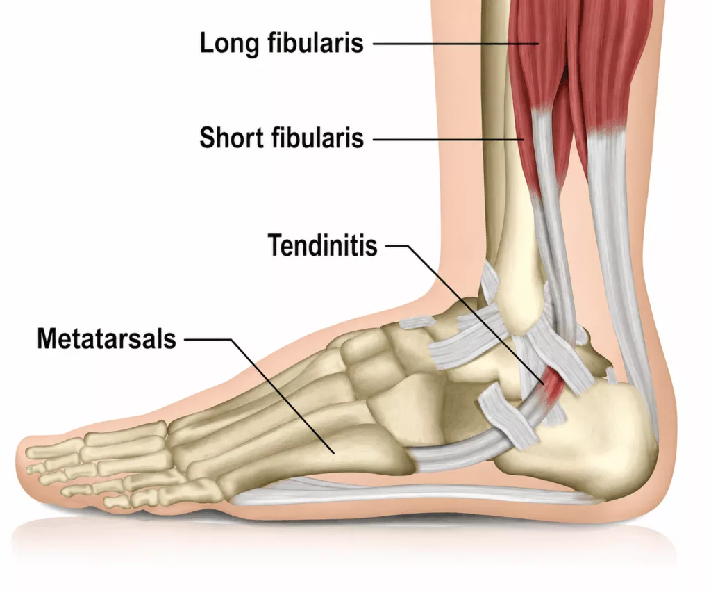
Two tendons run along the outer aspect of the ankle: the peroneus brevis and the peroneus longus tendons. They originate from the muscles of the lateral leg, pass through a groove behind the lateral malleolus, and insert distally onto the metatarsal bones. Their primary functions are to stabilise the ankle and facilitate plantarflexion and eversion of the foot.
Two main disorders affect the peroneal tendons:
- Peroneal tendinopathy, in which the tendons become inflamed, painful and progressively weakened, potentially leading to partial or complete tendon tears.
- Peroneal tendon instability (dislocation or subluxation), in which the tendons slip out of their normal groove behind the lateral malleolus.
Causes and Risk Factors for Peroneal Tendinopathy, Instability and Tendon Tears
Ankle Surgery Specialist in Hauts-de-Seine (92)
Several factors may contribute to the development of peroneal tendon disorders, including:
- recurrent ankle sprains and repeated microtrauma;
- anatomical variations affecting the bones or tendons;
- hindfoot varus deformity;
- inflammatory rheumatic diseases.
Peroneal Tendinopathy, Instability and Tendon Tears in Rueil-Malmaison
Ankle Surgery in Hauts-de-Seine
Symptoms of Peroneal Tendinopathy, Instability and Tendon Tears
Patients typically experience pain along the outer aspect of the ankle that worsens with physical activity and improves with rest. Local inflammation may also develop, with swelling and warmth around the tendons.
Symptoms are generally aggravated by ankle eversion and plantarflexion.
When the peroneal tendons become unstable or dislocate, patients often describe a snapping, popping or shifting sensation behind the lateral malleolus, frequently accompanied by feelings of ankle instability.
In cases of complete tendon rupture, pain may partially subside. However, patients usually develop persistent ankle instability with recurrent sprains and a noticeable loss of strength during foot eversion.
Diagnosis of Peroneal Tendinopathy, Instability and Tendon Tears
The consultation begins with a detailed medical history focusing on pain, swelling, local warmth within the retromalleolar groove and any sensations of snapping or clicking behind the lateral malleolus.
Clinical examination aims to reproduce symptoms by palpating the tendons and placing them under tension through resisted eversion and plantarflexion manoeuvres.
The strength of the peroneal tendons is also assessed, as weakness may indicate a partial or complete tendon tear.
Weight-bearing foot and ankle radiographs are obtained to evaluate the retromalleolar groove, assess overall foot alignment and identify any associated fractures or degenerative changes.
Magnetic resonance imaging (MRI) remains the reference investigation. It allows direct visualisation of the peroneal tendons, assessment of tendon continuity and identification of inflammatory changes such as bone marrow oedema or fluid within the peroneal tendon sheath.
Treatment of Peroneal Tendinopathy, Instability and Tendon Tears
Peroneal tendinopathy is initially managed conservatively with activity modification, anti-inflammatory medication and the use of a pronation-correcting orthotic in appropriate cases.
Corticosteroid injections are contraindicated, as they may weaken the tendons and increase the risk of rupture.
If symptoms persist despite conservative treatment, surgical management may be considered.
Complete rupture of the peroneal tendons requires surgical repair, as these tendons are unable to heal adequately without operative treatment.
Peroneal tendon instability or dislocation is also treated surgically. Simple immobilisation alone does not provide reliable long-term stabilisation of the tendons.