
Accessory Navicular Bone in Hauts-de-Seine

Foot Surgery

What is an accessory navicular bone?

Dr Simon Tournemine in Hauts-de-Seine (92)

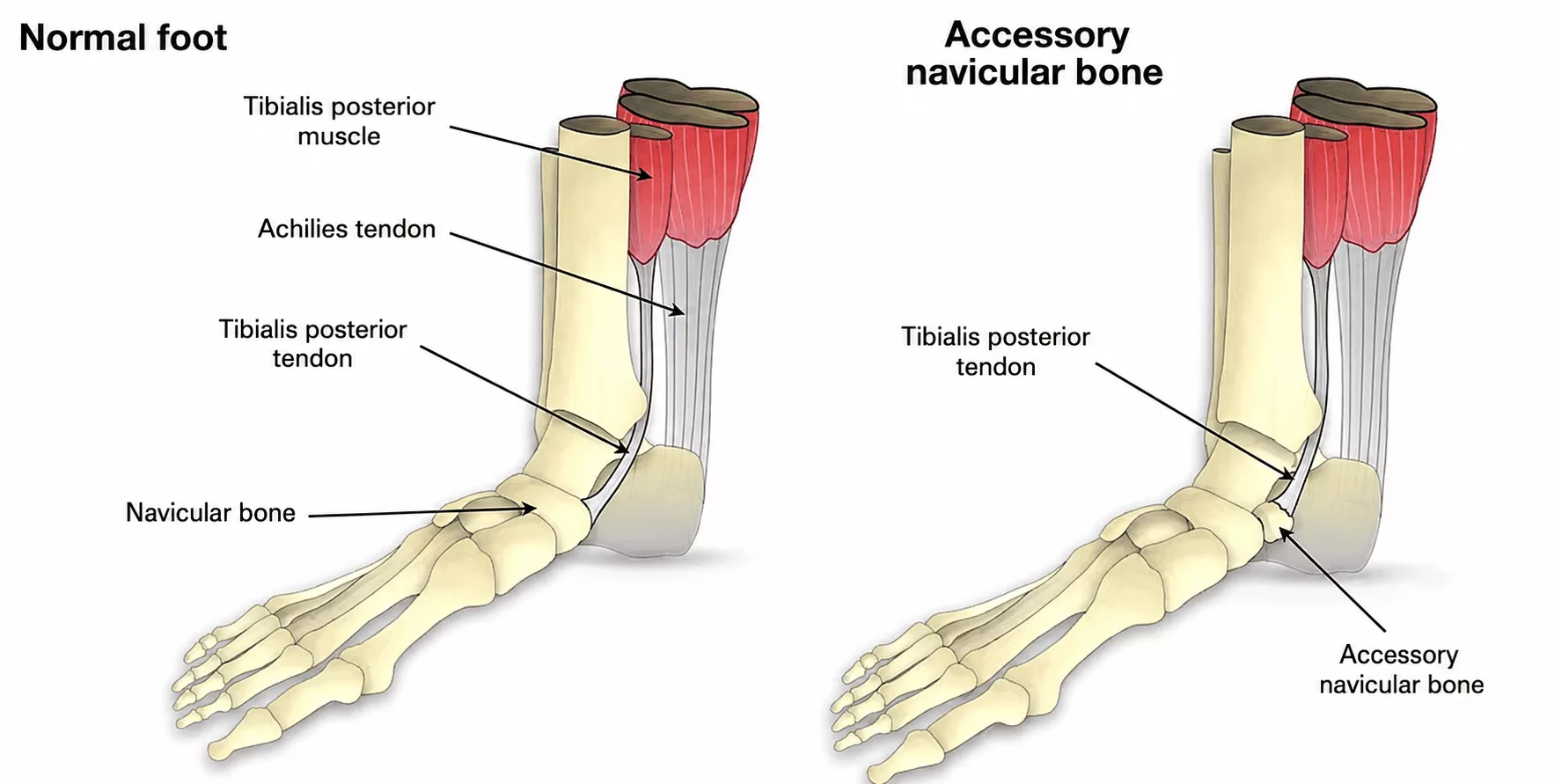
The navicular bone (tarsal navicular) is a central bone of the foot. It plays a key role in transmitting movement between the hindfoot and the forefoot. It also serves as the insertion site for the posterior tibial tendon, a powerful tendon responsible for plantarflexion and inversion of the foot.

Approximately 10% of the population has a normal anatomical variation in which a second, smaller navicular bone is connected to the main navicular by either a fibrous bridge (synfibrosis) or a cartilaginous bridge (synchondrosis). This is known as an accessory navicular bone. The posterior tibial tendon inserts onto this accessory bone.

Causes and risk factors for an accessory navicular bone

Specialist in Foot Surgery in Hauts-de-Seine (92)

The accessory navicular develops during skeletal growth. It is not a congenital malformation, but rather a common anatomical variant found in approximately 10% of the population.

It is also frequently bilateral, affecting both feet.

Accessory Navicular Bone in Rueil-Malmaison

Foot Surgery in Hauts-de-Seine

An accessory navicular typically presents as a painful bony prominence on the inner side of the foot, just below the ankle.

It may become painful during sporting activities or following excessive loading. Repetitive microtrauma can lead to inflammation at the fragile junction between the accessory navicular and the main navicular bone.

When the accessory bone is large, it may also rub against footwear, causing painful pressure and friction.

Over time, the accessory navicular may contribute to posterior tibial tendon insufficiency, leading to the gradual development of an adult-acquired flatfoot deformity.

Your consultation begins with a detailed medical history to identify factors contributing to the condition and to assess its impact on sporting activities and everyday life.

Clinical examination looks for a painful prominence on the inner aspect of the foot. Overall foot alignment, mobility and any associated deformities are also carefully assessed.

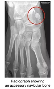
Weight-bearing anteroposterior and lateral foot radiographs are obtained to demonstrate the presence of the accessory navicular bone.

In atypical cases, magnetic resonance imaging (MRI) is required to identify inflammation between the main navicular and the accessory navicular, confirming that this anatomical variation is responsible for the patient’s symptoms.
In the majority of patients, conservative treatment is sufficient. This may include:

  • anti-inflammatory medication, either taken orally or applied locally as a cream to the painful area;
  • regular ice application during inflammatory flare-ups;
  • custom orthotic insoles with medial arch support to reduce stress on the posterior tibial tendon;
  • wearing soft, comfortable footwear.

Corticosteroid injections are not recommended, as they may weaken the posterior tibial tendon.

When symptoms persist despite appropriate conservative treatment, surgical management may be considered.

The procedure involves removing the accessory navicular bone and reattaching the posterior tibial tendon to the main navicular using either a bone anchor or an interference screw, depending on the surgical indication.