
Talo-Navicular Osteoarthritis in Hauts-de-Seine

Foot Surgery

What is talo-navicular osteoarthritis?

Dr Simon Tournemine in Hauts-de-Seine (92)

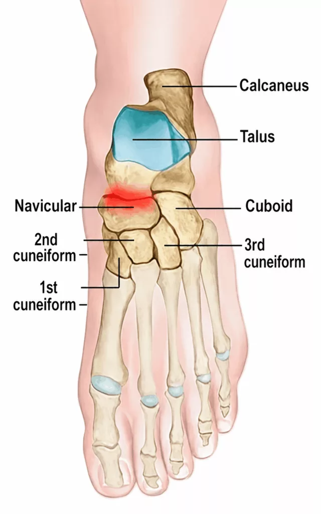
Talo-navicular osteoarthritis is a degenerative condition affecting the joint between the talus (ankle bone) and the navicular bone of the foot. Under normal circumstances, the joint surfaces are covered with a layer of smooth articular cartilage that allows the bones to glide painlessly over one another. Osteoarthritis develops when this cartilage progressively wears away.

The condition typically progresses in cycles, with:

  • chronic phases, during which pain remains relatively stable and moderate;
  • acute flare-ups characterised by significant pain and inflammation of the joint.

Talo-navicular osteoarthritis generally develops slowly over several years, resulting in progressively increasing discomfort and pain during walking and weight-bearing activities.

Degenerative joint disease is common, with approximately 80% of people over the age of 80 affected by osteoarthritis.

Causes and risk factors for talo-navicular osteoarthritis

Specialist in Foot Surgery in Hauts-de-Seine (92)

Talo-navicular osteoarthritis may be primary (without an identifiable anatomical or traumatic cause) or secondary, resulting from an underlying deformity or disease.

Primary osteoarthritis is associated with several recognised risk factors:

  • increasing age, with osteoarthritis becoming more common after the age of 60;
  • excess body weight;
  • female sex, particularly after the menopause.

Secondary osteoarthritis may develop as a result of:

  • congenital or acquired foot deformities, including flatfoot and high-arched foot (pes cavus);
  • recurrent ankle sprains;
  • fractures involving the talus or navicular bone;
  • inflammatory rheumatic diseases.

Talo-Navicular Osteoarthritis in Rueil-Malmaison

Foot Surgery in Hauts-de-Seine

Progressive pain is often the earliest symptom of talo-navicular osteoarthritis.

Pain is typically located over the dorsum of the foot, although it may radiate towards the ankle or toes. Symptoms worsen with walking and physical activity and are relieved by rest.

Patients frequently experience joint stiffness, particularly in the morning, requiring a short period of movement before the joint loosens.

If pain becomes severe or prolonged, a limp may develop and the use of a walking stick may become necessary.

The disease usually progresses slowly, alternating between painful flare-ups and periods of relative symptom relief.

Although osteoarthritis cannot always be prevented, several measures may help slow its progression and reduce symptoms.

Controlling modifiable risk factors through a healthy, balanced diet and appropriate footwear is recommended.

Maintaining a healthy body weight reduces stress across the joints and helps limit pain. It is equally important to protect the affected joint while remaining physically active. Low-impact exercises such as walking, cycling and swimming are particularly encouraged.

Your consultation begins with a detailed medical history to identify potential causes and risk factors for talo-navicular osteoarthritis. The impact of symptoms on daily life, including pain and difficulty wearing shoes, is also carefully assessed.

Clinical examination identifies the painful areas of the foot, evaluates foot alignment and mobility, and reproduces symptoms through mobilisation of the talo-navicular joint.

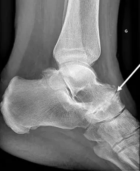
Weight-bearing anteroposterior and lateral radiographs confirm the diagnosis by demonstrating the characteristic radiographic signs of osteoarthritis:

  • narrowing of the joint space due to cartilage loss;
  • sclerosis of the underlying bone;
  • abnormal bony outgrowths around the joint known as osteophytes.

A computed tomography (CT) scan may also be required to evaluate the joint in greater detail and determine the severity of degenerative changes.
No treatment can reverse established talo-navicular osteoarthritis, but a range of therapeutic options can effectively relieve symptoms.

Initial management consists of conservative treatment, including:

  • weight reduction where appropriate;
  • maintaining regular physical activity;
  • pain relief with medications such as paracetamol and non-steroidal anti-inflammatory drugs (NSAIDs).

Intra-articular corticosteroid injections may also be offered to reduce pain and inflammation.

When conservative treatment no longer provides satisfactory symptom relief and daily activities remain significantly affected, surgical treatment may be recommended.

The surgical procedure consists of talo-navicular arthrodesis, in which the worn joint is fused to eliminate pain originating from the arthritic articulation.