
Accessory Navicular Excision in Hauts-de-Seine
Foot Surgery
What is accessory navicular excision?
Dr Simon Tournemine in Hauts-de-Seine (92)
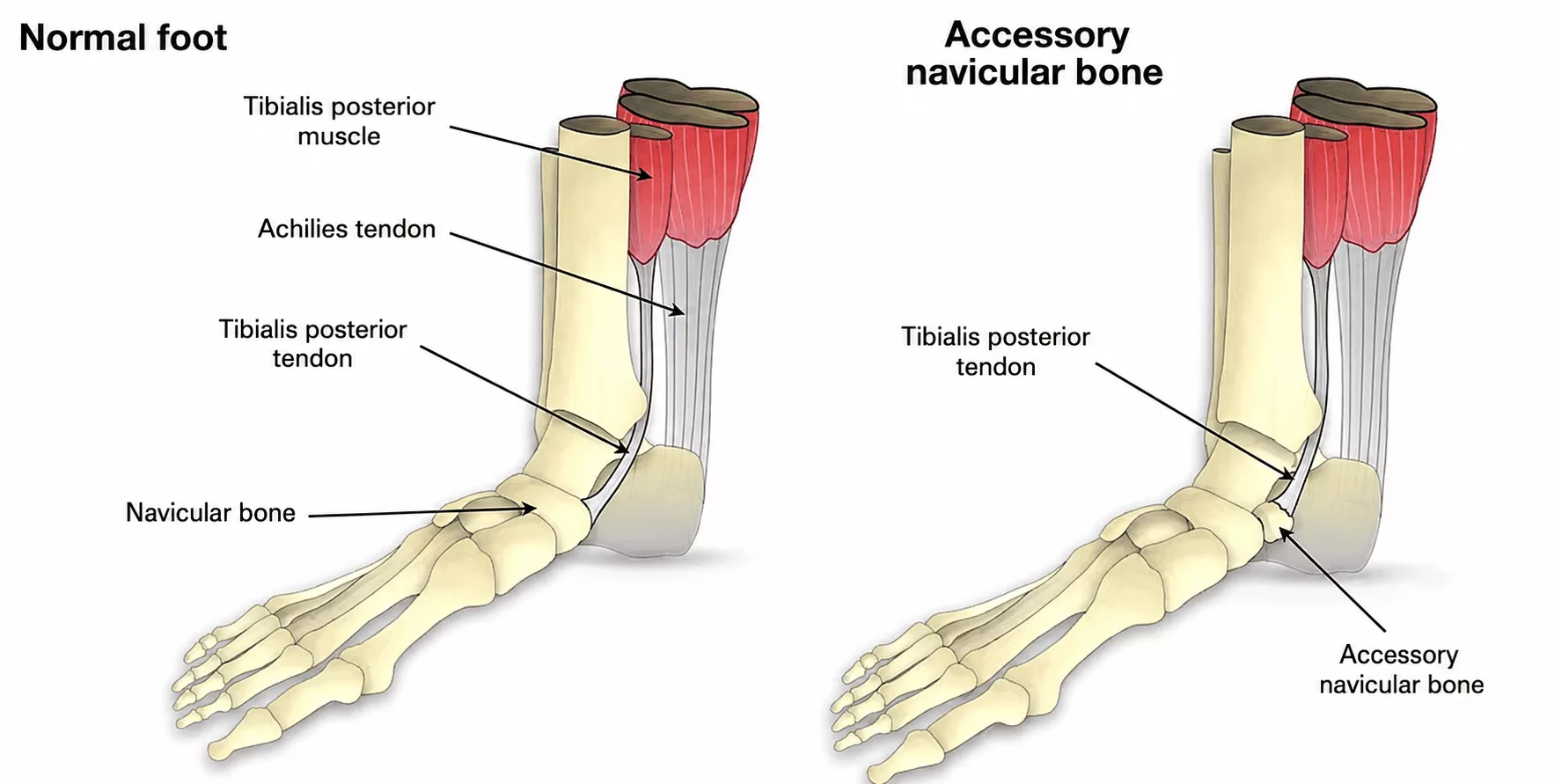
The navicular bone (also known as the tarsal scaphoid) is a central bone of the foot that plays an essential role in transmitting movement between the hindfoot and forefoot. It also serves as the insertion site for the posterior tibial tendon, a powerful tendon that supports the medial arch of the foot and contributes to plantar flexion and inversion.
Approximately 10% of the population have a common anatomical variation in which a second, smaller bone is connected to the main navicular by a fibrous bridge (synfibrosis) or a cartilaginous bridge (synchondrosis). This is known as an accessory navicular. In these cases, the posterior tibial tendon inserts onto the accessory bone rather than the main navicular.
The accessory navicular typically presents as a painful prominence on the inner side of the foot, just below the ankle. Symptoms often occur during sporting activities or periods of increased mechanical stress. Repetitive loading may cause inflammation at the fragile junction between the accessory navicular and the main navicular bone.
When the accessory bone is particularly large, it may also rub against footwear, causing pain and local irritation.
Over time, this anatomical variation may contribute to posterior tibial tendon dysfunction, potentially leading to the gradual development of an adult-acquired flatfoot deformity.
Accessory Navicular Excision
Specialist in Foot Surgery in Hauts-de-Seine (92)
A skin incision of approximately 5 cm is made along the inner side of the foot and ankle to expose the accessory navicular.
The accessory bone is removed completely.
As the posterior tibial tendon inserts onto the accessory navicular, it must first be detached before being securely reattached to the main navicular bone.
The tendon is fixed using either a suture anchor or a screw, providing strong fixation while healing occurs. This fixation device is generally well tolerated and does not usually require removal.
The procedure takes approximately 20 to 30 minutes.
It is performed under either general anaesthesia or regional anaesthesia (where only the leg is anaesthetised). The most appropriate anaesthetic technique will be discussed during your consultation with the anaesthetist.
The operation is performed as day-case (outpatient) surgery, allowing you to return home on the day of surgery.
Except in exceptional circumstances, both feet are not operated on simultaneously. An interval of approximately one month between procedures is generally recommended.
Accessory Navicular Excision in Rueil-Malmaison
Foot Surgery in Hauts-de-Seine
When should accessory navicular excision be performed?
In the majority of cases, conservative treatment is sufficient and includes:
- oral or topical anti-inflammatory medication;
- regular icing during painful inflammatory episodes;
- custom orthotic insoles supporting the medial arch to reduce stress on the posterior tibial tendon;
- soft, comfortable footwear;
- avoidance of corticosteroid injections, which are generally not recommended because they may weaken the tendon.
When symptoms persist despite appropriate conservative treatment, surgical treatment may be recommended.
Postoperative care and rehabilitation following accessory navicular excision
The procedure is performed as day-case surgery, allowing you to return home on the day of the operation.
Pain management is closely supervised to maximise comfort and facilitate recovery.
Anticoagulant treatment is prescribed throughout the immobilisation period to reduce the risk of deep vein thrombosis.
The foot and ankle are immobilised for four weeks to allow healing of the tendon repair.
During this period, walking is only permitted without weight-bearing using two crutches.
After approximately one month, the cast is removed and progressive weight-bearing begins using a removable walking boot.
Driving is generally possible after six weeks, while sporting activities can usually resume after approximately three months.
Physiotherapy begins once immobilisation has been discontinued in order to restore muscle strength, mobility and normal foot function.
These recovery times represent average estimates and may vary according to individual healing.
Regular clinical and radiographic follow-up is recommended to monitor healing and detect any complications.
Risks and complications of accessory navicular excision
Every precaution is taken to ensure optimal healing. Nevertheless, as with any surgical procedure, complications may occur, although they remain uncommon:
- deep vein thrombosis (DVT) and pulmonary embolism, with the risk reduced by anticoagulant treatment;
- worsening of pre-existing medical conditions such as heart disease or diabetes, monitored by the anaesthetic team;
- infection (risk below 1%). Should infection occur, further surgery to clean the operative site together with antibiotic treatment may be required;
- haematoma, which may occasionally require drainage or blood transfusion;
- injury to small sensory nerve branches supplying the ankle and foot, potentially resulting in areas of reduced sensation;
- Complex Regional Pain Syndrome (CRPS), a painful inflammatory condition that remains poorly understood. It is managed medically and may require prolonged rehabilitation, further investigations and specialised pain management. Both its occurrence and long-term outcome remain unpredictable;
- hardware-related irritation, where the implanted fixation device causes discomfort or irritation of surrounding tissues. In rare cases, removal of the implant may subsequently be required.
These represent the principal potential complications, although this list is not exhaustive. Rare or exceptional complications may also occur. Your surgeon will discuss your individual situation in detail during your consultation.
Results of accessory navicular excision
The objectives of surgery are to:
- relieve pain and restore comfortable walking and footwear;
- prevent skin complications such as calluses, blisters, ulcers and infection;
- reduce the long-term risk of developing progressive flatfoot deformity associated with posterior tibial tendon insufficiency.
Complete healing and return to sporting activities are generally achieved approximately 12 weeks after surgery.