
Osteochondral Lesions of the Talus (OLT) in Hauts-de-Seine
Ankle Surgery
What Are Osteochondral Lesions of the Talus (OLT)?
Dr Simon Tournemine in Hauts-de-Seine (92)
A joint is a complex structure that enables smooth movement between two bones. To allow friction-free, pain-free motion, the articulating bone surfaces are covered with articular cartilage.
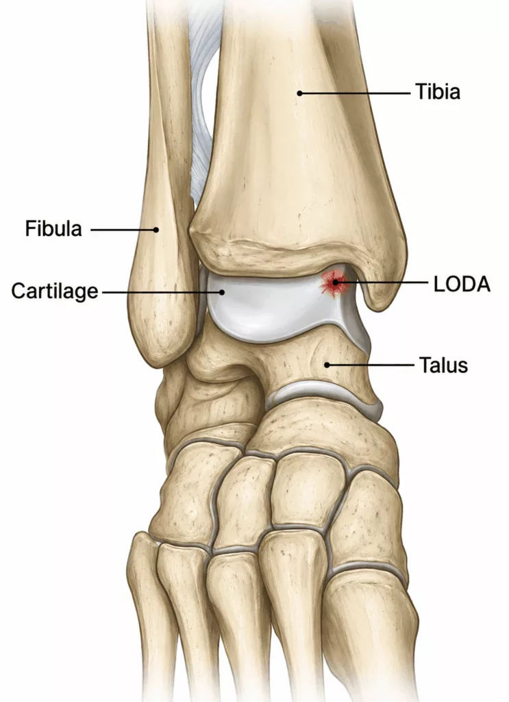
An osteochondral lesion of the talus (OLT) is an injury affecting both the cartilage and the underlying bone of the talar dome, the upper portion of the talus that articulates with the tibia to form the ankle joint.
Causes and Risk Factors for Osteochondral Lesions of the Talus
Ankle Surgery Specialist in Hauts-de-Seine (92)
There are three main types of osteochondral lesions of the talus:
- osteochondral fractures;
- osteonecrotic lesions;
- subchondral cysts.
Osteochondral Fractures
These lesions are typically located on the lateral aspect of the talar dome and most commonly occur following an ankle sprain.
Patients may experience persistent ankle pain, mechanical locking or a sensation of instability.
Treatment most often consists of arthroscopic removal of the detached fragment. When the fragment is sufficiently large and viable, it may be repositioned and securely fixed back into place.
Osteonecrotic Lesions
These lesions are generally located on the medial side of the talar dome and are frequently discovered incidentally during imaging.
A small area of the subchondral bone gradually loses its blood supply, causing bone collapse and secondary damage to the overlying cartilage.
Treatment usually involves arthroscopic debridement and curettage of the lesion. In selected cases, an osteochondral autograft transplantation (mosaicplasty), harvested from the patient’s knee, may be recommended to restore the damaged cartilage and underlying bone.
Subchondral Cysts
Subchondral cysts correspond to cavities within the talus while the overlying cartilage often remains relatively preserved.
They may develop following previous trauma, although in many cases no specific cause can be identified.
When these cysts are responsible for persistent pain, treatment consists of curettage followed by bone grafting, performed either arthroscopically or through an open surgical approach depending on the location and size of the lesion.
Osteochondral Lesions of the Talus in Rueil-Malmaison
Ankle Surgery in Hauts-de-Seine
Symptoms of Osteochondral Lesions of the Talus
Gradually progressive ankle pain is often the earliest symptom. Pain typically worsens with physical activity and weight-bearing and improves with rest.
Symptoms frequently fluctuate, with painful episodes alternating with periods of relative improvement.
Patients may also experience recurrent ankle swelling caused by joint effusion.
When loose osteochondral fragments are present within the joint, mechanical symptoms such as clicking, catching or locking may occur as these fragments become trapped between the articular surfaces.
Diagnosis of Osteochondral Lesions of the Talus
The consultation begins with a detailed medical history to identify possible causes of the osteochondral lesion and assess symptoms including pain, clicking, locking and their impact on daily activities.
Clinical examination focuses on identifying areas of tenderness around the ankle. The surgeon also evaluates joint effusion, ankle range of motion and ligament stability to detect any associated ligament injuries.
Imaging investigations are routinely performed.
Standard ankle radiographs may reveal the lesion and associated cartilage degeneration in some cases.
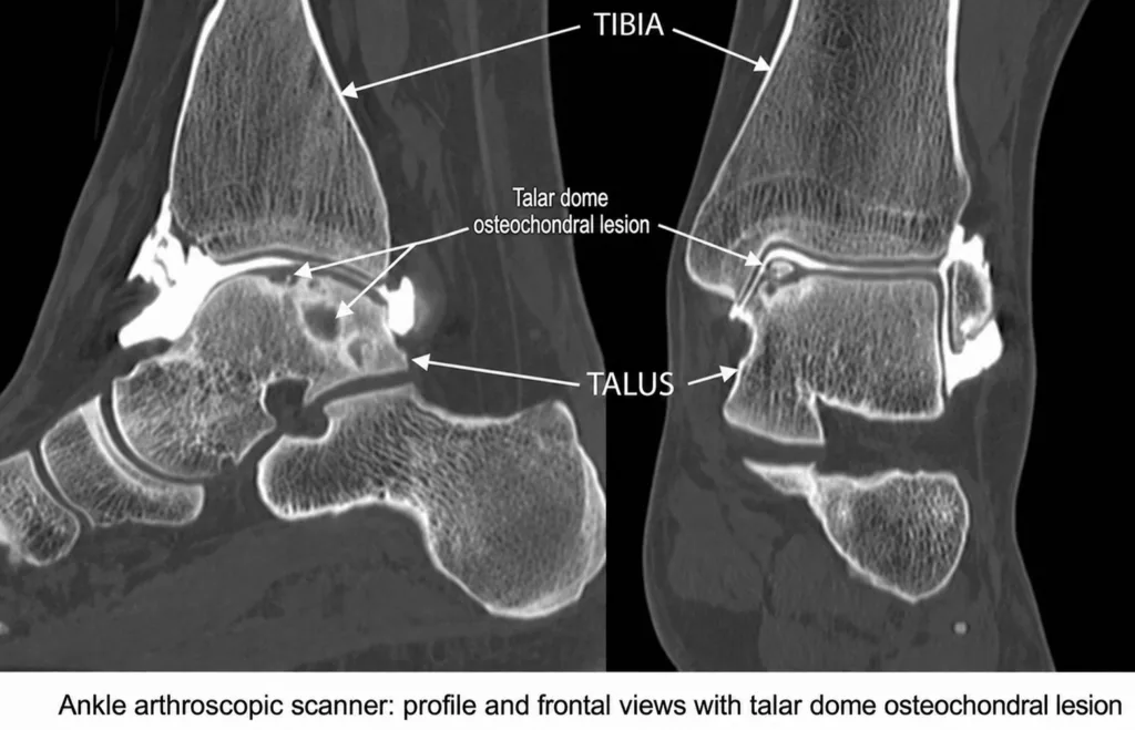
Magnetic resonance imaging (MRI) and CT arthrography remain the reference imaging modalities, allowing precise assessment of the osteochondral lesion, the presence of loose fragments, cartilage damage and lesion size. These findings guide treatment planning. Imaging also helps identify any associated ligament injuries.
Treatment of Osteochondral Lesions of the Talus
Management of osteochondral lesions is highly individualised.
Treatment depends on several factors, including:
- the severity of symptoms;
- the type of lesion;
- its size and depth;
- the presence of loose osteochondral fragments.
Several surgical techniques are available, including:
- microfracture surgery;
- osteochondral grafting (mosaicplasty);
- fixation of the detached osteochondral fragment.
The most appropriate treatment is determined individually following careful assessment and discussion with your surgeon.