
Synostosis and Synchondrosis in Hauts-de-Seine
Foot Surgery
What are synostosis and synchondrosis?
Dr Simon Tournemine in Hauts-de-Seine (92)
Synostosis and synchondrosis are anatomical abnormalities of the foot characterised by the persistence of abnormal connections between certain foot bones. These bridges may be bony (synostosis) or cartilaginous (synchondrosis) and restrict the normal movement that should exist between the affected bones.
Synostoses may occur between any of the bones of the foot. However, talocalcaneal and calcaneonavicular coalitions account for approximately 90% of all diagnosed cases, while other forms are considerably less common.
Causes and risk factors for synostosis and synchondrosis
Specialist in Foot Surgery in Hauts-de-Seine (92)
During childhood, much of the foot skeleton is composed of cartilage, which gradually ossifies between the ages of 10 and 13 years.
During this developmental process, some bones may form an abnormal bony bridge instead of developing a normal joint that allows movement between them.
In some patients, these abnormal bridges may develop following a fracture. However, in the majority of cases, no specific cause can be identified.
Synostosis and Synchondrosis in Rueil-Malmaison
Foot Surgery in Hauts-de-Seine
Symptoms of synostosis and synchondrosis
Because the abnormal bridge joins two bones together, normal foot movement becomes restricted, which may lead to:
- pain directly over the affected coalition;
- a feeling of instability when walking on uneven ground;
- a flat foot deformity;
- pain during foot movement;
- recurrent ankle sprains.
Diagnosis of synostosis and synchondrosis
Your consultation begins with a detailed medical history to assess the nature of your symptoms and their impact on daily activities.
Clinical examination looks for several characteristic findings, including a precisely localised painful area within the foot, reduced mobility of the midfoot and hindfoot, pain reproduced during rotational stress testing, a flat foot deformity, and evidence of previous ankle sprains.
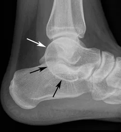
Weight-bearing anteroposterior and lateral foot radiographs are obtained to assess overall foot alignment, investigate other potential causes of pain, evaluate any sequelae of previous ankle sprains, and, in some cases, directly demonstrate the coalition.
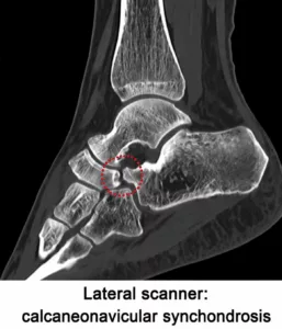
Computed tomography (CT) and magnetic resonance imaging (MRI) are the reference imaging examinations. They accurately define the location, type and extent of the synostosis or synchondrosis, information that plays an important role in planning the most appropriate treatment.
Treatment of synostosis and synchondrosis
There are many different types of synostosis and synchondrosis, and every patient has different functional demands. For this reason, treatment should always be tailored individually and discussed with your surgeon.
Many coalitions are discovered incidentally and cause no symptoms. In these cases, no treatment is required.
When the coalition becomes symptomatic, initial management consists of conservative treatment, including custom orthotic insoles, activity modification, physiotherapy and anti-inflammatory medication.
If symptoms persist despite appropriate non-surgical treatment, surgical management may be recommended.
Depending on the type of coalition and several patient-specific factors, surgery may involve either:
- resection of the coalition, removing the abnormal bony bridge while preserving joint motion; or
- arthrodesis, in which the painful malformed joint is fused.
The most appropriate surgical option depends on multiple factors and is determined on an individual basis in consultation with your surgeon.