Heel spur removal in the Hauts-de-Seine department

Foot Surgery

What is a heel spur resection?

Dr Simon Tournemine in Hauts-de-Seine (92)

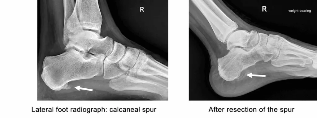
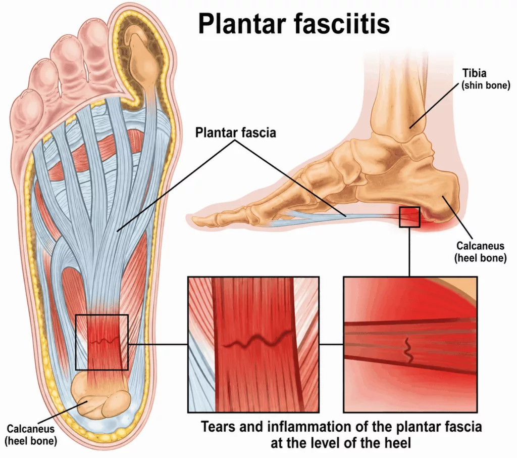
A heel spur is a bony growth located beneath the heel, on the calcaneus, at the junction with the plantar fascia. It develops as a result of inflammation of the plantar fascia, a ligament connecting the heel to the toes. When the plantar fascia is subjected to excessive trauma and stretching, inflammation develops, leading to calcification.

A heel spur is often caused by a combination of factors such as excessive strain on the heel, poor foot posture (flat feet or high arches), sports activities that put strain on the feet, or being overweight. It most commonly occurs between the ages of 40 and 60 and is more prevalent in women.

It is characterised by sharp pain under the heel. This pain is felt when taking the first steps upon waking, or after standing for a prolonged period.

Surgery to remove the heel spur

Specialist in Foot Surgery in Hauts-de-Seine (92)

The surgery involves removing the heel spur and releasing the plantar fascia by cutting through the middle two-thirds. This is combined with the release of the lateral plantar nerve, which is often compressed at the heel. To do this, a 4–5 cm incision is made on the inner side of the heel.

The procedure takes 30 minutes. It is carried out under general or regional anaesthesia (only the leg is numbed). You will decide on the type of anaesthesia in consultation with the anaesthetist during your consultation.

The operation is carried out on an outpatient basis; you will be able to go home on the evening of the operation.

You will be allowed to walk again on the same day, wearing a removable walking boot for three weeks, with partial weight-bearing for the first two weeks.

Heel spur removal in Rueil-Malmaison

Foot Surgery in Hauts-de-Seine

A simple course of medical treatment is initially put in place. This consists of weight loss, limiting activities that put strain on the foot, and wearing suitable footwear (shoes with flat, flexible soles). Orthopaedic insoles with shock-absorbing heel cups are also prescribed, along with physiotherapy involving massage and stretching of the plantar fascia, and shockwave therapy. In cases of severe pain, anti-inflammatory treatment or corticosteroid injections may be used on an ad hoc basis.

If medical treatment proves ineffective, surgical treatment is considered.

A sterile dressing is applied in the operating theatre and must be left in place for two weeks without being removed. Pain management is initiated and closely monitored to ensure maximum comfort and a speedy recovery.

Anticoagulant treatment is required for two weeks to thin the blood and reduce the risk of phlebitis.

You may resume walking on the same day, wearing a removable walking boot for three weeks, two of which should be spent on partial weight-bearing.

After the operation, the foot and toes will be swollen (oedema); this is normal. To help the oedema subside more quickly, rest and keep your foot elevated for the first two weeks.

You will then need to undertake self-rehabilitation with daily stretching exercises for the gastrocnemius and hamstring muscles.

Sick leave is generally for two weeks, but this depends on your occupation. Office work can usually be resumed more quickly. You may resume sporting activities after six weeks. These timeframes are averages; they vary from person to person and are provided for guidance only.

Subsequently, regular clinical and X-ray follow-up is essential to monitor your foot and detect any abnormalities.

Every measure has been taken to ensure a favourable outcome. However, there is a risk of surgical complications, which are rare but possible:

  • phlebitis and pulmonary embolism: a rare complication, prevented by prescribing anticoagulants to thin the blood for two weeks;
  • infection (risk less than 1 per cent). In this case, a further operation is required to clean the surgical site, followed by a course of antibiotics;
  • haematoma: it is possible that the operated area may bleed after the procedure and form a haematoma. Depending on the extent of the bleeding, it may be necessary to drain the haematoma;
  • Persistent pain: whilst surgery reduces pain in the majority of cases, some residual post-operative pain may persist.
  • Algodystrophy is a painful and inflammatory condition that is still not fully understood. It is treated medically and can last for several months (or sometimes even years), requiring specific management with appropriate rehabilitation, further investigations and, in some cases, specialised pain management. Its onset, progression and potential long-term effects are unpredictable.

These are the main risks, but the list is not exhaustive. Other rare complications may occur. It is not possible to list all complications, a fact which you understand and accept. Your surgeon is available to discuss each individual case with you.

Without treatment, plantar fasciitis tends to take a negative course, with the pain worsening.

In 90 per cent of cases, medical treatment is sufficient, but if the pain persists, surgery may be necessary. The aim is to reduce or even eliminate the pain so that the patient can resume their normal activities and walk normally.