Cartilage Surgery in Hauts-de-Seine
Knee Surgery
What Is Cartilage Surgery?
Dr Simon Tournemine in Hauts-de-Seine (92)
A joint is a complex anatomical structure that allows movement between two bones. To enable the bones to glide smoothly and without pain, their surfaces are covered with articular cartilage.
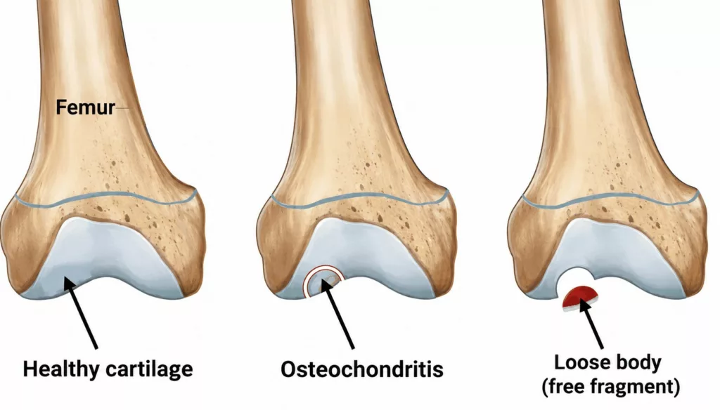
Osteochondritis is a lesion affecting both the articular cartilage and the underlying bone. A small area of bone supporting the cartilage loses its vitality, gradually becomes weaker and eventually collapses, taking the overlying cartilage with it. Bone fragments may then progressively detach and become loose within the joint.
The onset of knee pain is usually the first symptom. It may be accompanied by recurrent episodes of swelling caused by joint effusion. When loose fragments are present within the knee, patients may also experience clicking, catching or episodes of mechanical locking.
Cartilage Surgery Procedure
Knee Surgery Specialist in Hauts-de-Seine (92)
The management of cartilage lesions is complex. Treatment depends on several factors, including the severity of the patient’s symptoms, the size and depth of the lesion, the presence of loose fragments and the overall degree of joint wear.
Several treatment options may be considered, including:
- Microfracture surgery
- Mosaicplasty using an osteochondral graft
- Fixation of a detached fragment
- Knee replacement as a last resort
Treatment must be tailored to each patient and discussed on an individual basis with the orthopaedic surgeon.
Cartilage Surgery in Rueil-Malmaison
Knee Surgery in Hauts-de-Seine
Cartilage Surgery Procedure
Microfracture Surgery
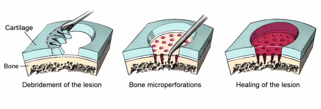
This technique aims to stimulate bone and cartilage repair by creating small perforations, several millimetres deep, in the exposed bone beneath the cartilage lesion.
These perforations encourage a healing response from the underlying bone, leading to the formation of fibrocartilage over the damaged surface.
Mosaicplasty
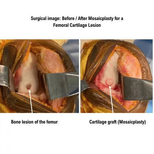
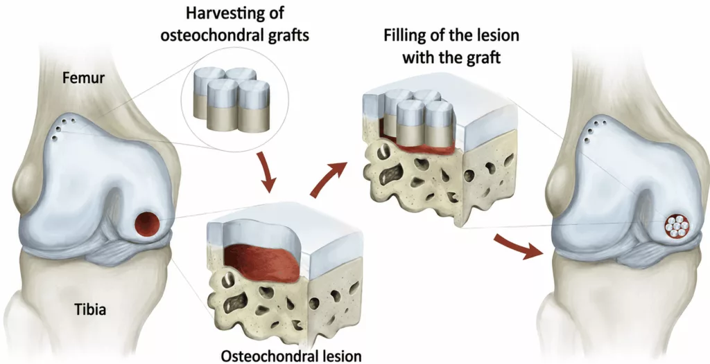
Mosaicplasty consists of replacing the damaged bone and cartilage with an osteochondral graft.
A cylindrical plug of healthy bone and cartilage, corresponding to the size of the lesion, is harvested from a non-weight-bearing area of the femoral trochlea. It is then implanted into the damaged area.
The graft subsequently integrates with the surrounding bone and recreates a cartilage-covered joint surface. The donor site is not normally a source of pain or wear because it is located within a part of the joint that is not subjected to significant mechanical loading.
Fixation of a Detached Fragment
When a large osteochondral fragment becomes detached, it may be repositioned anatomically and secured using buried screws.
The fragment can then heal in its original position and restore the normal anatomy of the joint surface.
Knee Replacement
When the lesion is too extensive and cartilage wear is advanced, the previous cartilage-preserving procedures may no longer be sufficient. In this situation, the damaged joint surface may be replaced with a unicompartmental knee replacement.
Whenever possible, these procedures are performed arthroscopically. In some cases, however, it may be necessary to open the knee.
Arthroscopy is a minimally invasive, video-assisted technique that avoids opening the joint. Two incisions measuring approximately 5 mm are made at the front of the knee to introduce a small camera and the instruments required to treat the lesion.
This technique offers several advantages, including a more discreet scar, less postoperative pain and faster recovery.
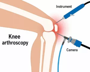
These procedures generally last approximately 45 minutes.
They are performed under either general anaesthesia or spinal anaesthesia, in which only the lower limbs are anaesthetised. The choice of anaesthetic is made jointly with the anaesthetist during the preoperative consultation.
The operation is usually performed as day-case surgery, allowing patients to return home on the same day.
Postoperative Care and Rehabilitation After Cartilage Surgery
The dressing is generally changed every two days for approximately two weeks. Pain management is closely monitored to maximise comfort and facilitate early rehabilitation.
Anticoagulant treatment is prescribed for six weeks to reduce the risk of deep vein thrombosis.
Physiotherapy begins immediately after surgery to preserve muscle strength and maintain knee flexibility.
Following mosaicplasty or fixation of a loose osteochondral fragment, the operated knee must be protected from full weight-bearing while the bone heals. Patients therefore walk with two crutches and reduced weight-bearing for approximately six weeks.
Driving is generally possible after six weeks, and sporting activities may usually be resumed after three months.
Following microfracture surgery, the period of reduced weight-bearing may be limited to approximately three weeks. Driving may then be resumed after three weeks, while sporting activities may generally restart after six weeks.
These timeframes are averages and are provided for guidance only, as recovery varies from one patient to another.
Long-term clinical and radiographic follow-up remains essential to monitor the knee and identify any potential abnormalities.
When Is Cartilage Surgery Recommended?
A cartilage lesion is initially managed with conservative treatment. This includes resting the knee and taking anti-inflammatory medication. Corticosteroid injections may also be prescribed.
When conservative treatment does not provide sufficient improvement and the patient continues to experience pain and limitations in everyday or sporting activities, surgical treatment may be considered.
Why Undergo Cartilage Surgery? What Are the Benefits?
The natural progression of a cartilage lesion is generally unfavourable. Articular cartilage cannot heal spontaneously, and the lesion may therefore gradually increase in size, progressively damaging the entire joint and eventually leading to osteoarthritis.
Cartilage surgery has two principal objectives:
- To relieve pain and allow patients to return to normal daily and sporting activities
- To preserve the knee and limit long-term joint wear and the progression of osteoarthritis
Risks and Complications of Cartilage Surgery
Every precaution is taken to promote a favourable recovery. Nevertheless, as with any surgical procedure, complications, although uncommon, may occur.
The principal risks include:
- Deep vein thrombosis and pulmonary embolism: uncommon complications, with the risk reduced by anticoagulant treatment prescribed after surgery;
- Worsening of a pre-existing medical condition: such as heart failure or diabetes. Postoperative monitoring by the anaesthetic team is provided to reduce this risk;
- Infection, with a risk below 1%: this may require further surgery to wash out the surgical site, followed by antibiotic treatment;
- Reduced knee mobility and postoperative stiffness: these may develop if rehabilitation is not carried out appropriately;
- Haematoma: postoperative bleeding may lead to the formation of a haematoma. Depending on its severity, drainage or blood transfusion may be required;
- Sensory nerve injury: small sensory nerve branches around the knee may rarely be injured, resulting in reduced sensation over certain areas of the knee;
- Complex Regional Pain Syndrome (CRPS): a painful and inflammatory condition that remains poorly understood. It is treated medically and may persist for several months or, in some cases, several years. It may require adapted rehabilitation, additional investigations and specific pain management. Its onset, progression and possible long-term consequences are unpredictable.
These are the principal risks associated with cartilage surgery, but the list is not exhaustive. Other exceptional complications may occur. Not every possible complication can be specified. Your surgeon remains available to discuss the particular risks associated with your individual situation.
Results of Cartilage Surgery
Following fixation of an osteochondral fragment, bone healing is achieved in approximately 75% to 90% of cases, with traumatic lesions generally showing a higher healing rate.
Following microfracture surgery, pain and mechanical locking improve in approximately 80% of patients when the cartilage lesion is isolated.
Following mosaicplasty, good pain-related outcomes are achieved in approximately 90% of cases. Minor discomfort at the graft donor site may occur in approximately 15% of patients.
Regardless of the procedure performed, significant improvement is achieved in the majority of cases, with recovery of knee strength and mobility generally occurring within two to three months.