
Anterior Ankle Impingement in Hauts-de-Seine

Ankle Surgery

What Is Anterior Ankle Impingement?

Dr Simon Tournemine in Hauts-de-Seine (92)

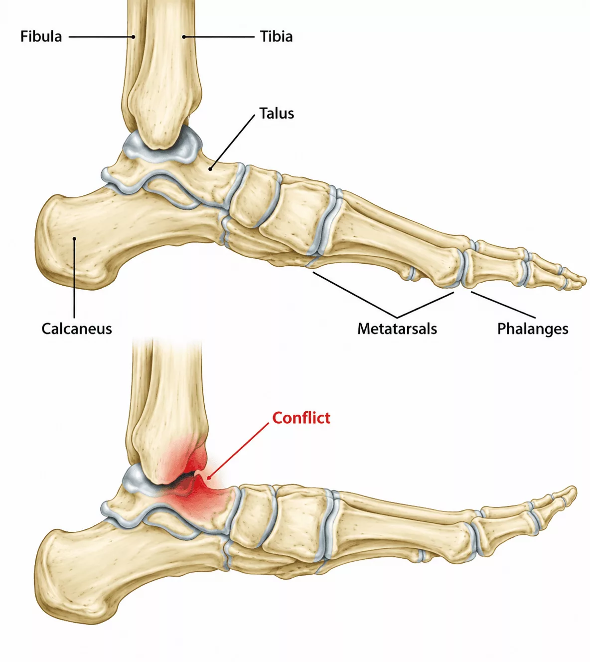
The ankle joint forms the articulation between the shin bone (tibia) and the talus. It allows the foot to move upwards (dorsiflexion) and downwards (plantarflexion). An ankle impingement occurs when structures become trapped within the joint during movement, causing pain and restricting motion. These impinging structures may consist of bony outgrowths (osteophytes) or soft tissues such as ligaments or the synovial membrane.

Two main types of ankle impingement are recognised:

  • Anterior ankle impingement, when the impinging structures are located at the front of the ankle;
  • Posterior ankle impingement, when the impingement occurs at the back of the ankle.

Causes and Risk Factors for Anterior Ankle Impingement

Ankle Surgery Specialist in Hauts-de-Seine (92)

Anterior ankle impingement most commonly develops following repeated ankle sprains or recurrent ankle trauma.

These injuries damage the ligaments and adjacent bone. During healing, excessive scar tissue, ligament thickening and the formation of bony osteophytes may occur. These enlarged soft tissues and bony prominences can become trapped within the joint during ankle motion, leading to painful pinching and mechanical restriction.

Anterior Ankle Impingement in Rueil-Malmaison

Ankle Surgery in Hauts-de-Seine

Anterior ankle impingement typically presents as pain at the front of the ankle during physical activity or movements requiring ankle dorsiflexion.

The precise location of the pain often helps identify the underlying cause:

  • anterolateral pain generally suggests soft tissue impingement;
  • anteromedial pain is more commonly associated with bony impingement caused by osteophytes.

Pain is frequently accompanied by a sensation of ankle instability resulting from ligament damage sustained during previous sprains.

Without appropriate treatment, repeated impingement may progressively damage the joint cartilage and ultimately contribute to the development of ankle osteoarthritis.

The consultation begins with a detailed medical history to identify the underlying cause of the impingement and assess symptoms such as pain, clicking, locking and their impact on daily activities.

Weight-bearing radiographs of the foot and ankle help identify bony impingement, evaluate cartilage wear, assess overall foot alignment and detect any associated bony abnormalities.

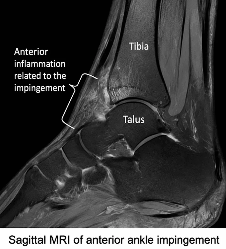
CT arthrography and magnetic resonance imaging (MRI) are the reference imaging investigations. They provide detailed visualisation of the osteophytes and soft tissue lesions responsible for the impingement while also assessing the condition of the ankle ligaments and articular cartilage.
Initial management is conservative.

Treatment typically combines temporary activity modification with anti-inflammatory medication. In selected patients, a corticosteroid injection into the impingement site may provide effective symptom relief.

When conservative treatment fails to relieve pain or restore function, surgical treatment may be recommended.

The procedure is usually performed arthroscopically and involves removing the bony osteophytes and inflamed soft tissues responsible for the impingement, thereby restoring joint mobility and relieving pain.