Cartilage Surgery & Osteochondral Lesions of the Talus (OLT) in Hauts-de-Seine

Ankle Surgery

What is an Osteochondral Lesion of the Talus (OLT)?

Dr Simon Tournemine in Hauts-de-Seine (92)

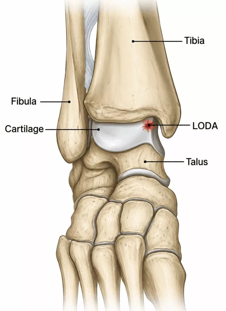
A joint is a complex structure that enables smooth movement between two bones. To allow pain-free motion with minimal friction, the bony surfaces are covered by articular cartilage. An osteochondral lesion of the talus (OLT) is an injury affecting both this cartilage and the underlying bone of the talar dome, which forms part of the ankle joint.

There are three main types of OLT:

  • Fracture
  • Osteonecrosis
  • Subchondral cyst

Types of Osteochondral Lesions of the Talus (OLT)

Specialist in Ankle Surgery in Hauts-de-Seine (92)

Fractures

These lesions are located on the lateral aspect of the talar dome and are most commonly associated with an ankle sprain. They may cause pain, episodes of locking or catching, and a feeling of ankle instability.

Treatment most often consists of arthroscopic removal of the detached fragment. When the fragment is sufficiently large and viable, it may instead be repositioned and securely fixed.

Osteonecrosis

These lesions are usually located on the medial side of the talar dome and are often discovered incidentally.

A small area of bone beneath the cartilage loses its blood supply and gradually weakens. As the bone collapses, the overlying cartilage also deteriorates.

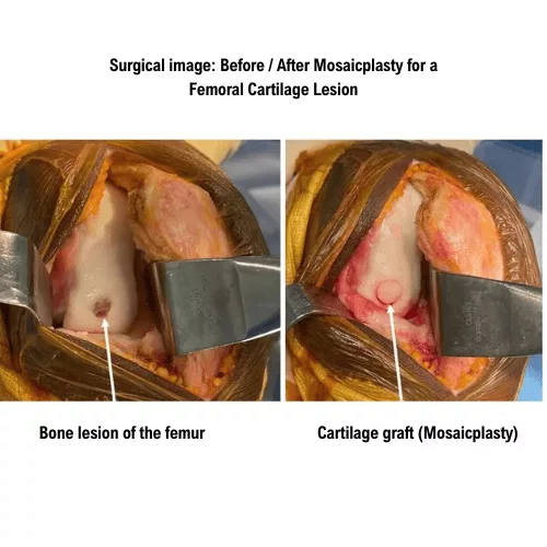
Treatment involves arthroscopic debridement and curettage of the lesion. In some cases, an osteochondral graft (mosaicplasty) harvested from the knee may be required.

Subchondral cysts

These lesions correspond to cavities within the talus while the overlying cartilage often remains relatively preserved.

They may occasionally develop following previous trauma, although in many cases no definite cause can be identified.

When they are responsible for pain, treatment consists of curettage and bone grafting, performed either arthroscopically or through an open procedure.

Treatment should always be individualised and discussed on a case-by-case basis with your surgeon.

Cartilage Surgery & OLT in Rueil-Malmaison

Ankle Surgery in Hauts-de-Seine

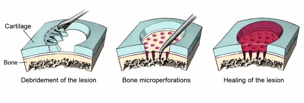
Curettage and microfracture

This technique stimulates bone healing by first removing the damaged tissue and then creating multiple small perforations in the exposed subchondral bone. These microfractures promote the formation of new bone and repair cartilage.
Bone grafting

Bone grafting involves filling the defect using bone harvested from the knee. A cylindrical bone graft matching the size of the lesion is removed and transplanted into the defect.

The graft subsequently consolidates and restores normal anatomy. This procedure does not produce significant consequences at the donor site, as the harvested bone heals naturally over time.

Fragment fixation

When a large osteochondral fragment becomes detached, it may be repositioned anatomically and fixed using buried screws. Once healed, the fragment restores the normal architecture of the joint surface.

Whenever possible, these procedures are performed arthroscopically. However, in certain cases, an open operation may be required.

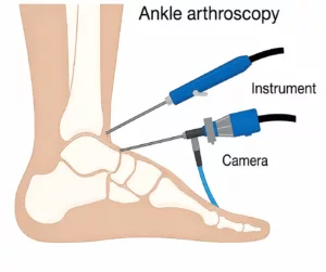
Arthroscopy is a minimally invasive, video-assisted technique that avoids opening the ankle joint. Two small incisions of approximately 10 mm are made to introduce a miniature camera together with the specialised instruments required to repair the lesion.

Compared with traditional open surgery, arthroscopy offers several advantages, including reduced blood loss, less postoperative pain, smaller scars and faster recovery.
These procedures take approximately 30 minutes.

They are performed under either general anaesthesia or spinal anaesthesia (where only the legs are anaesthetised). The most appropriate anaesthetic technique will be discussed with your anaesthetist during your preoperative consultation.

The operation is performed as day-case surgery, allowing you to return home on the same day.

Dressings should be changed every two days for approximately two weeks, while the wound heals.

Pain management is carefully monitored to maximise comfort and facilitate early rehabilitation. Anticoagulant treatment is prescribed during the period of immobilisation to reduce the risk of deep vein thrombosis.

Physiotherapy should begin immediately to preserve muscle strength and maintain ankle mobility.

Following bone grafting or refixation of a detached fragment, the ankle must remain partially unloaded using two crutches for approximately six weeks to allow bone healing. Driving is generally possible after six weeks, and sporting activities may usually be resumed after approximately three months.

Following curettage and microfracture, the protected weight-bearing period may be reduced to approximately three weeks. Driving is generally possible after three weeks, while sporting activities may be resumed after around six weeks.

These recovery periods are average estimates and vary according to each patient’s individual progress.

Long-term clinical and radiographic follow-up remains essential to monitor the ankle and detect any potential abnormalities.

An OLT is initially managed conservatively with temporary rest, anti-inflammatory medication and, in some cases, corticosteroid injections.

If conservative treatment fails to relieve symptoms and pain continues to limit daily activities, surgical treatment may be recommended.

Every precaution is taken to ensure the safest possible outcome. Nevertheless, as with any surgical procedure, complications, although uncommon, may occur.

Potential complications include:

  • Deep vein thrombosis (DVT) and pulmonary embolism: uncommon complications minimised through anticoagulant treatment during the postoperative period.
  • Exacerbation of pre-existing medical conditions: such as heart failure or diabetes. Careful postoperative monitoring is provided by the anaesthetic team.
  • Infection (risk below 1%): this may require further surgery to clean the operative site, followed by antibiotic treatment.
  • Reduced ankle mobility and postoperative stiffness: these may occur if rehabilitation is not carried out appropriately.
  • Haematoma: postoperative bleeding may occasionally result in a haematoma requiring drainage or, more rarely, blood transfusion.
  • Injury to small superficial sensory nerve branches: this uncommon complication may lead to reduced sensation in certain areas around the ankle.
  • Complex Regional Pain Syndrome (CRPS): a rare and poorly understood painful inflammatory condition that may persist for several months, or occasionally years, requiring specialised rehabilitation, additional investigations and, in some cases, dedicated pain management. Its onset, progression and long-term consequences remain unpredictable.

These represent the principal recognised risks but do not constitute an exhaustive list. Other rare or exceptional complications may occur. Not every possible complication can be listed, and your surgeon will be pleased to discuss any specific concerns relating to your individual case.

The natural progression of osteochondral cartilage lesions is unfavourable. Articular cartilage has very limited healing potential, meaning that untreated lesions gradually enlarge, eventually affecting the entire joint and leading to ankle osteoarthritis.

The objectives of surgery are twofold:

  • to relieve pain;
  • to preserve the ankle joint and reduce long-term degenerative changes, including osteoarthritis.

Surgical outcomes vary according to the type of osteochondral lesion.

The best results are generally achieved in fracture-type OLTs. Outcomes are also favourable for small osteonecrotic lesions and small subchondral cysts.

Conversely, results may be less predictable for large or deep osteochondral defects. In these situations, conservative management with corticosteroid injections may be preferred, while more extensive surgical procedures, such as ankle arthrodesis, may ultimately become necessary.