
Heel Spur and Plantar Fasciitis in Hauts-de-Seine

Foot Surgery

What is a heel spur?

Dr Simon Tournemine in Hauts-de-Seine (92)

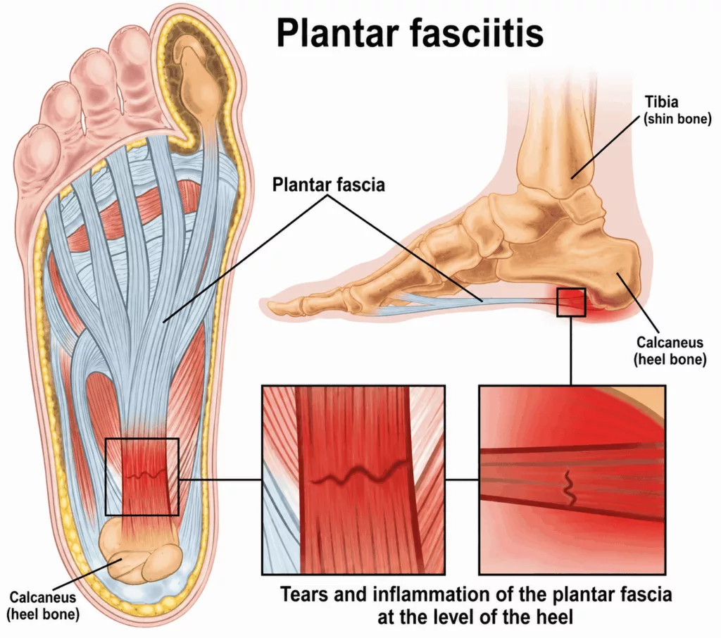
A heel spur (calcaneal spur) is a bony outgrowth that develops beneath the heel bone (calcaneus), at the insertion of the plantar fascia. Its formation results from chronic inflammation of the plantar fascia, a strong fibrous band that extends from the heel to the toes and supports the longitudinal arch of the foot.

When the plantar fascia is subjected to repeated mechanical stress and excessive traction, inflammation develops at its attachment to the calcaneus. Over time, this chronic inflammatory process may lead to the formation of a calcified bony spur.

Causes and risk factors for a heel spur

Specialist in Foot Surgery in Hauts-de-Seine (92)

Heel spurs usually result from a combination of contributing factors, including excessive loading of the heel, abnormal foot biomechanics such as flatfoot (pes planus) or high-arched foot (pes cavus), participation in high-impact sports, and excess body weight.

The condition most commonly affects adults between 40 and 60 years of age and is more frequently observed in women.

Heel Spur and Plantar Fasciitis in Rueil-Malmaison

Foot Surgery in Hauts-de-Seine

Several measures can help reduce inflammation around the heel and limit the development or progression of a heel spur:

  • maintaining a healthy body weight;
  • wearing flat shoes with cushioned soles;
  • performing regular plantar fascia stretching exercises;
  • limiting activities that place repetitive impact on the heel.

A heel spur is typically associated with sharp pain beneath the heel.

Pain is often most intense during the first steps taken in the morning or after prolonged standing. Symptoms are primarily related to inflammation of the plantar fascia, which may also cause local swelling (oedema) and warmth.

The consultation begins with a detailed medical history to identify the potential causes and risk factors associated with heel spurs. The impact of symptoms on everyday activities is also carefully assessed.

Clinical examination identifies the painful areas and evaluates the overall alignment and biomechanics of the foot. Direct palpation over the plantar aspect of the heel typically reproduces the patient’s pain.

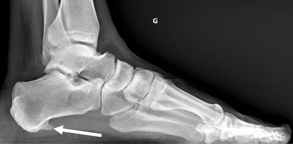
Weight-bearing anteroposterior and lateral radiographs are obtained to visualise the calcaneal spur and exclude other potential causes of heel pain.

Magnetic resonance imaging (MRI) remains the reference investigation for assessing inflammation of the plantar fascia and excluding alternative diagnoses.
Initial management is based on conservative treatment.

This includes:

  • weight reduction where appropriate;
  • limiting activities that place repetitive stress on the heel;
  • adapting footwear by choosing flat shoes with soft, cushioned soles;
  • custom-made orthotic insoles incorporating a shock-absorbing heel pad;
  • physiotherapy with massage techniques and stretching exercises targeting the plantar fascia;
  • extracorporeal shockwave therapy.

During acute painful episodes, anti-inflammatory medication may also be prescribed.

If conservative treatment fails to provide satisfactory relief and symptoms persist, surgical treatment may be considered.