
An accessory navicular bone is a common anatomical variant found in approximately 15% of the population. Although it is usually asymptomatic, this extra bone located along the inner side of the foot can sometimes become a source of chronic pain and discomfort.
When symptoms develop, one important question arises: should surgery be considered, or can conservative treatment provide sufficient relief?
This article explains when surgery may be appropriate and reviews the most effective non-surgical treatment options.
What is an accessory navicular bone?
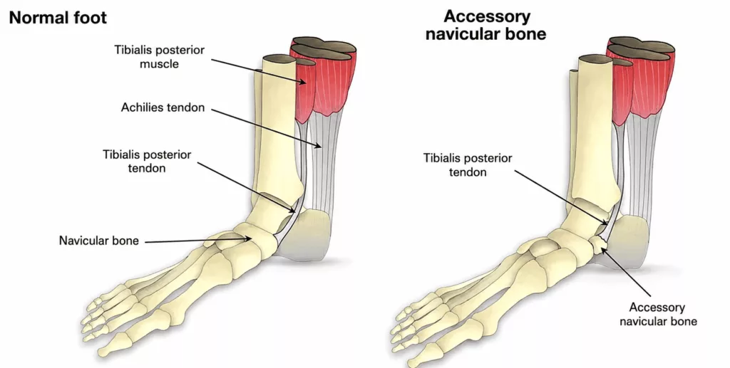
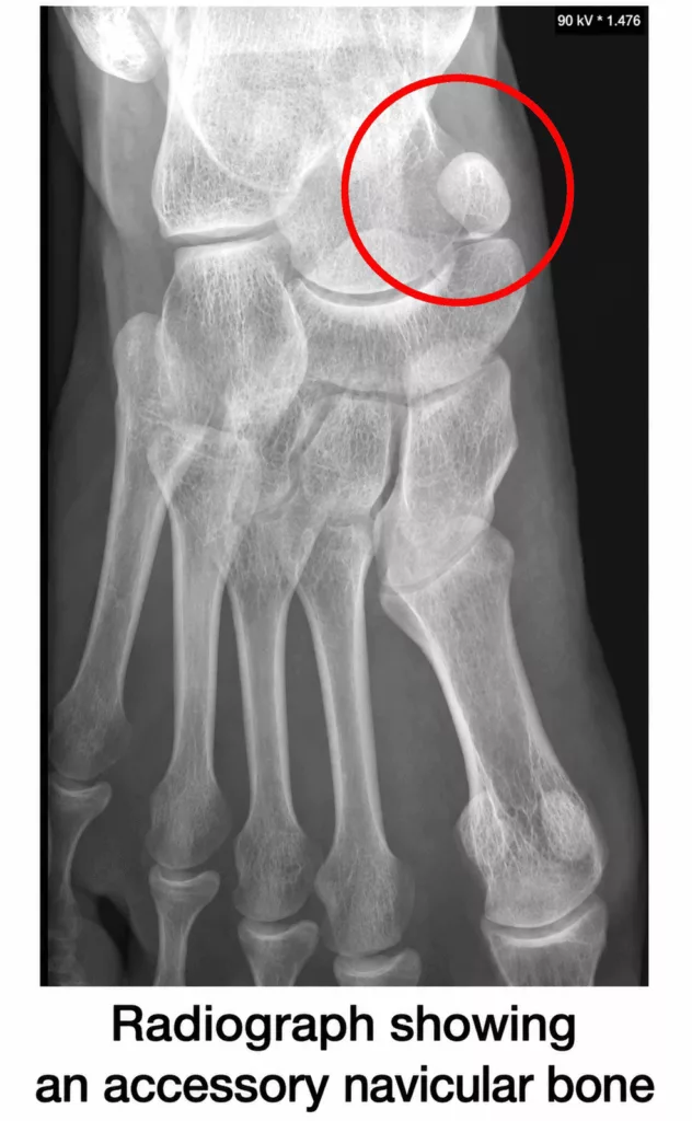
An accessory navicular is a small extra bone located adjacent to the navicular bone on the inner aspect of the foot, within the medial arch.
It is embedded within the posterior tibial tendon, which plays a vital role in supporting the arch of the foot and maintaining normal foot mechanics.
Most people are unaware that they have an accessory navicular until symptoms are triggered by an ankle sprain, repetitive sporting activities or pressure from unsuitable footwear. Inflammation may then develop, causing a visible bony prominence, local redness and pain during movement or weight-bearing.
Alternatives to surgery: First-line treatment
Surgery is not the initial treatment of choice. Conservative management aims to reduce inflammation and decrease the mechanical stress placed on the posterior tibial tendon.
The principal non-surgical treatment options include:
1. Rest and activity modification
The first step is to reduce or temporarily avoid activities that aggravate symptoms, particularly running and impact sports.
During acute flare-ups, temporary offloading with crutches or immobilisation in a walking boot may be recommended to allow the inflamed tissues to settle.
2. Ice therapy and anti-inflammatory medication
Applying ice several times a day helps reduce swelling and discomfort.
When appropriate, non-steroidal anti-inflammatory drugs (NSAIDs), either oral or topical, may also be prescribed to control pain and inflammation.
3. Custom orthotic insoles
This is often the most effective long-term treatment.
Custom-made orthoses support the medial arch and help correct flatfoot (pes planovalgus), which is frequently associated with a symptomatic accessory navicular.
By supporting the arch, orthoses reduce excessive traction on the posterior tibial tendon, thereby relieving mechanical irritation around the accessory bone.
4. Physiotherapy
A structured rehabilitation programme strengthens the posterior tibial muscle and the intrinsic muscles of the foot.
Physiotherapy also focuses on improving ankle flexibility, balance and lower limb biomechanics in order to reduce the risk of symptom recurrence.
5. Corticosteroid injection
If symptoms persist despite conservative treatment, a local corticosteroid injection may be considered.
This can provide effective short-term relief by reducing inflammation around the affected tissues, although its benefits are generally temporary.
When should an accessory navicular be treated surgically?
Surgery is considered only after an adequate trial of conservative treatment has failed.
In most cases, an operation is discussed after three to six months of well-conducted non-operative management without significant improvement.
The main indications for surgery include:
- Persistent disabling pain that limits sporting activities or causes discomfort during everyday walking.
- Failure of orthotic treatment, with ongoing inflammation despite regular use of custom insoles.
- Difficulty wearing footwear, when the bony prominence causes persistent pressure and painful rubbing inside shoes.
What does the operation involve?
The most commonly performed procedure is the Kidner procedure.
This operation is usually carried out as day-case surgery, allowing patients to return home on the same day.
The procedure involves:
- removal of the accessory navicular bone;
- reshaping of the prominent portion of the native navicular bone to reduce future irritation;
- reattachment of the posterior tibial tendon to the remaining navicular bone in order to restore its important role in supporting the medial arch.
Following surgery, the foot is generally immobilised in a cast for approximately six weeks, followed by progressive physiotherapy.
Return to sporting activities is usually possible after approximately three months, depending on healing and functional recovery.
Conclusion
An accessory navicular bone is a common anatomical variation that does not usually require surgery.
For the majority of symptomatic patients, a combination of rest, appropriately fitted orthotic insoles and physiotherapy provides effective relief.
Surgery offers excellent outcomes but is reserved for patients with persistent symptoms that fail to respond to conservative treatment and significantly affect quality of life.
If you are experiencing pain along the inner side of your foot, early assessment by a foot and ankle orthopaedic surgeon is recommended to establish an accurate diagnosis and determine the treatment most appropriate for your condition.