
Achilles Tendon Rupture in Hauts-de-Seine
Ankle Surgery
What Is an Achilles Tendon Rupture?
Dr Simon Tournemine in Hauts-de-Seine (92)
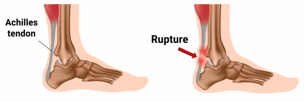
The Achilles tendon connects the calf muscles (triceps surae) to the heel bone (calcaneus). When the calf muscles contract, they pull on the tendon, producing plantarflexion of the ankle and providing the propulsion required for walking, running and jumping.
Although the Achilles tendon is the strongest tendon in the human body, it can rupture following a sudden and forceful contraction of the calf muscles, most commonly during sporting activities such as tennis, football or athletics. It may also rupture following excessive stretching of the tendon during a fall.
As we age, the tendon gradually becomes less resistant and may rupture after relatively minor trauma, such as a simple misstep.
Most ruptures occur within the mid-portion of the tendon, although some occur at its insertion into the calcaneus.
Causes and Risk Factors for an Achilles Tendon Rupture
Ankle Surgery Specialist in Hauts-de-Seine (92)
Achilles tendon ruptures occur most frequently in physically active men over the age of 40.
Certain medications can weaken the tendon and increase the risk of rupture, particularly fluoroquinolone antibiotics and corticosteroid injections.
Other recognised risk factors include:
- chronic Achilles tendinopathy;
- excess body weight;
- repetitive tendon overuse.
Achilles Tendon Rupture in Rueil-Malmaison
Ankle Surgery in Hauts-de-Seine
Preventing an Achilles Tendon Rupture
Regular physical activity helps maintain strong tendons and healthy muscles.
Before participating in sport, it is important to perform an appropriate warm-up, stretch adequately, remain well hydrated and wear footwear suited to the activity.
Symptoms of an Achilles Tendon Rupture
Patients often describe hearing or feeling a sudden snap or tearing sensation in the heel at the moment of injury.
Pain then develops in the lower calf and heel and may radiate into the leg. Bruising subsequently appears around the site of the rupture.
Standing on tiptoe becomes impossible because the tendon is no longer able to transmit force from the calf muscles.
Although walking usually remains possible, it is painful and associated with a marked loss of ankle strength. This preserved ability to walk frequently leads patients to underestimate the injury and may delay specialist assessment.
Diagnosis of an Achilles Tendon Rupture
The consultation begins by identifying potential risk factors and establishing the circumstances surrounding the injury.
Clinical examination looks for several characteristic signs, including:
- tenderness and bruising along the tendon;
- a palpable gap at the site of rupture;
- loss of the normal resting plantarflexed position of the foot (loss of physiological equinus);
- a positive Thompson test, demonstrated by the absence of ankle plantarflexion when the calf muscle is squeezed;
- inability to stand on the toes of the injured foot.
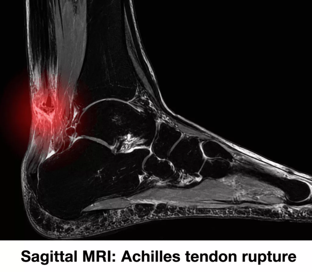
Magnetic resonance imaging (MRI) or ultrasound is performed to confirm the diagnosis, determine the exact location of the rupture and assess its extent. These findings are important in selecting the most appropriate treatment.
Treatment of an Achilles Tendon Rupture
An untreated Achilles tendon rupture results in permanent loss of tendon function.
If the tendon is not repaired promptly, the torn ends progressively retract and are no longer able to heal together. Although walking generally remains possible, patients experience significant weakness of the calf muscles and are usually unable to run or participate in sporting activities.
Two treatment options are available:
- Conservative treatment, consisting of immobilisation in a plaster cast or walking boot with the ankle held in an equinus position;
- Surgical treatment, which consists of repairing the tendon by suturing the torn ends together.
The choice between conservative and surgical treatment depends on several factors, including the patient’s age, sporting level, general health and associated medical conditions. The most appropriate management should be discussed individually with your orthopaedic surgeon.