
Haglund's Deformity in Hauts-de-Seine

Foot Surgery

What is Haglund's deformity?

Dr Simon Tournemine in Hauts-de-Seine (92)

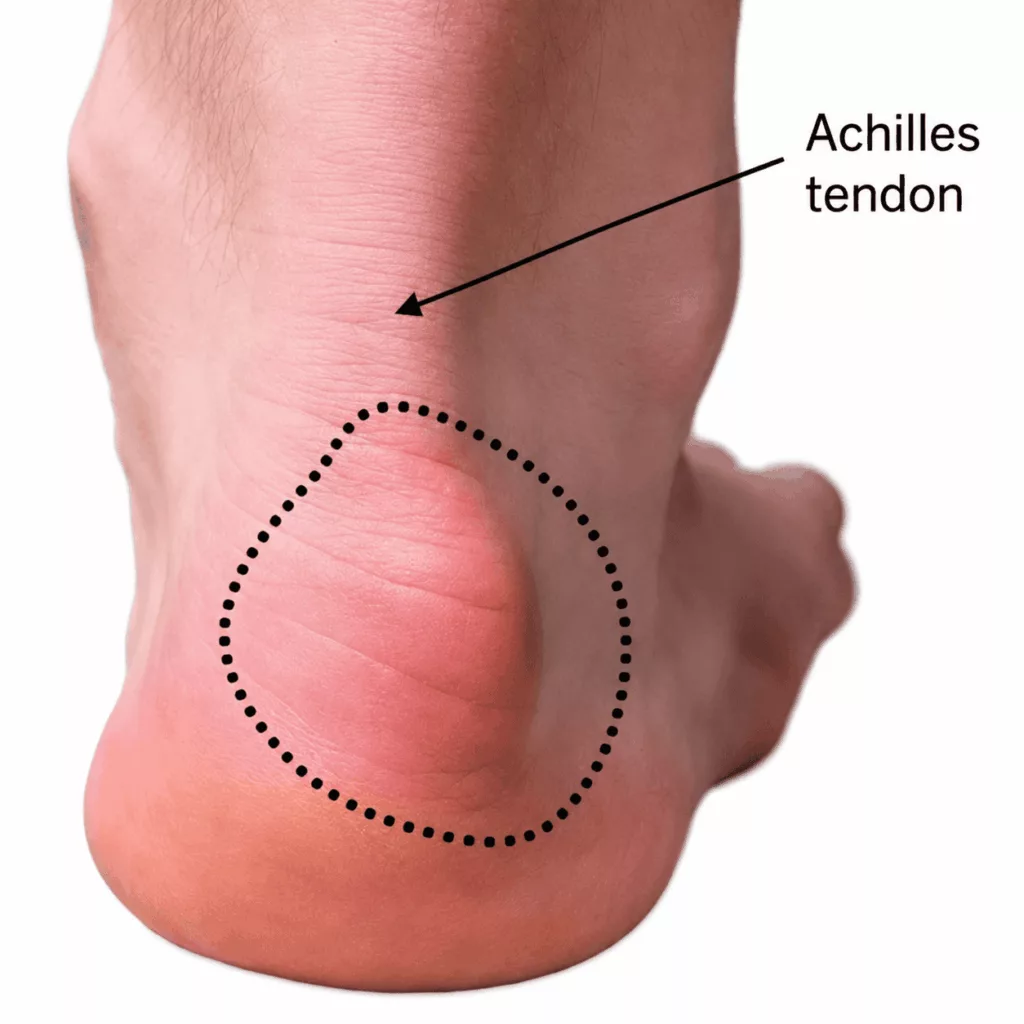
Haglund’s deformity is an Achilles insertional enthesopathy, meaning a disorder affecting the point where the Achilles tendon attaches to the heel bone (calcaneus).

It is characterised by the development of a bony prominence at the back of the heel, associated with pain and inflammation at the insertion of the Achilles tendon. The condition often causes discomfort when walking, exercising or wearing certain types of footwear due to repeated friction over the affected area.

Causes and risk factors for Haglund's deformity

Specialist in Foot Surgery in Hauts-de-Seine (92)

Haglund’s deformity is often bilateral and is most commonly diagnosed in adults between 20 and 30 years of age.

Several factors are known to increase the risk of developing the condition, including:

  • shoes with rigid heel counters (such as safety footwear);
  • high-heeled shoes;
  • a high-arched foot (pes cavus).

Haglund's Deformity in Rueil-Malmaison

Foot Surgery in Hauts-de-Seine

The main symptoms include:

  • pain at the back of the heel during walking and sporting activities;
  • a visible bony prominence at the back of the heel;
  • swelling and redness around the heel;
  • corns (areas of thickened skin) or blisters developing where the heel rubs against footwear.

Your consultation begins with a detailed medical history to identify the factors contributing to the condition and to assess its impact on sporting activities and everyday life.

Clinical examination looks for a painful, prominent swelling at the insertion of the Achilles tendon on the heel. The overall alignment of the foot is assessed, together with ankle mobility and stability, in order to identify any associated conditions.

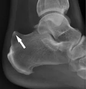
Weight-bearing lateral ankle radiographs are obtained to evaluate the size of the posterior calcaneal prominence and to identify any calcification at the Achilles tendon insertion. The overall morphology of the foot, including the presence of a high-arched or flat foot, is also assessed to help guide treatment.

Magnetic resonance imaging (MRI) remains the reference examination, allowing direct visualisation of the tendon lesion, its extent and any associated abnormalities.
In the majority of patients, conservative treatment provides satisfactory symptom relief. This may include:

  • anti-inflammatory medication, either taken orally or applied locally as a cream to the painful area;
  • regular ice application during inflammatory flare-ups;
  • physiotherapy including massage and stretching exercises targeting the Achilles tendon;
  • custom orthotic insoles with a heel lift to reduce tension on the Achilles tendon and minimise friction within the shoe;
  • wearing shoes with a soft heel counter.

Corticosteroid injections are not recommended, as they may weaken the Achilles tendon and increase the risk of tendon injury.

When symptoms persist despite appropriate non-surgical treatment, surgical management may be considered.