
Knee Osteochondritis in Hauts-de-Seine

Knee Surgery

What Is Knee Osteochondritis?

Dr Simon Tournemine in Hauts-de-Seine (92)

The knee is a complex joint that enables smooth, pain-free movement between the femur, tibia and patella. The ends of these bones are covered with articular cartilage, allowing the joint surfaces to glide smoothly during movement.

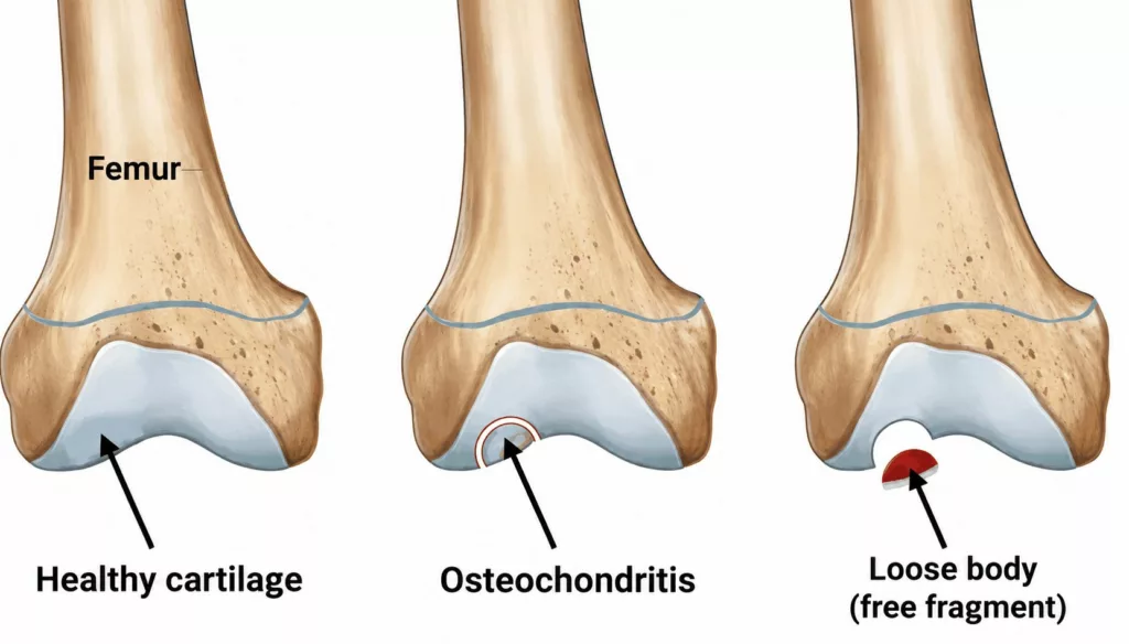
Knee osteochondritis is a condition affecting both the articular cartilage and the underlying bone (subchondral bone). A small area of bone beneath the cartilage gradually loses its blood supply and structural strength, eventually collapsing and causing the overlying cartilage to deteriorate. As the condition progresses, fragments of bone and cartilage may gradually detach and become loose within the knee joint, leading to mechanical symptoms.

Causes and Risk Factors for Knee Osteochondritis

Knee Surgery Specialist in Hauts-de-Seine (92)

Knee osteochondritis most commonly affects young, physically active individuals. Men are affected approximately twice as often as women.

In many cases, the cartilage lesion develops following a clearly identifiable traumatic injury or as a result of repeated microtrauma to the knee. Previous knee surgery and the long-term consequences of a fracture are also recognised risk factors.

However, despite thorough investigation, no specific cause can be identified in a significant number of patients.

Knee Osteochondritis in Rueil-Malmaison

Knee Surgery in Hauts-de-Seine

Gradually increasing knee pain is often the first symptom of knee osteochondritis. The pain typically worsens during physical activity and improves with rest. Symptoms usually fluctuate over time, with painful flare-ups alternating with periods of relative improvement. Episodes of knee swelling caused by joint effusion are also common.

When loose bone or cartilage fragments are present within the joint, patients may experience clicking sensations, catching or episodes of knee locking as these fragments become trapped between the joint surfaces.

The consultation begins with a detailed medical history to establish the possible cause of the osteochondral lesion and to assess the patient’s symptoms, including pain, clicking sensations and episodes of locking. The type of sport practised and the patient’s level of physical activity are also taken into consideration.

Clinical examination includes careful palpation and assessment of knee movement to identify painful areas. The presence of a joint effusion is evaluated, and knee stability is assessed to identify any associated ligament or meniscal injuries.

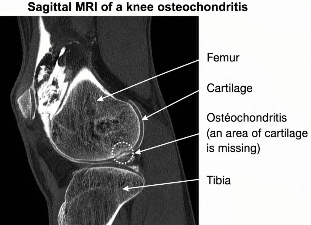
Imaging studies are systematically performed alongside the clinical examination. Standard knee X-rays help identify the osteochondral lesion and assess cartilage degeneration. Magnetic resonance imaging (MRI) is the reference investigation, providing detailed visualisation of the osteochondral defect, the condition of the articular cartilage and any loose fragments within the joint. MRI also allows accurate measurement of the size of the lesion, which is essential for selecting the most appropriate treatment. In addition, it evaluates associated injuries involving the menisci or knee ligaments.
The management of knee osteochondritis is often complex and must be tailored to each individual patient. Treatment depends on several factors, including the severity of symptoms, the size and depth of the lesion, the patient’s level of sporting activity and the presence of loose osteochondral fragments within the joint.

Several surgical techniques may be considered, including:

  • Microfracture surgery
  • Mosaicplasty (osteochondral grafting)
  • Osteochondral fragment fixation

The most appropriate treatment is determined on a case-by-case basis following a detailed discussion with your orthopaedic surgeon.