
Meniscal Surgery in the Hauts-de-Seine
Knee Surgery
What is meniscal surgery?
Dr Tournemine | Hauts-de-Seine (92)
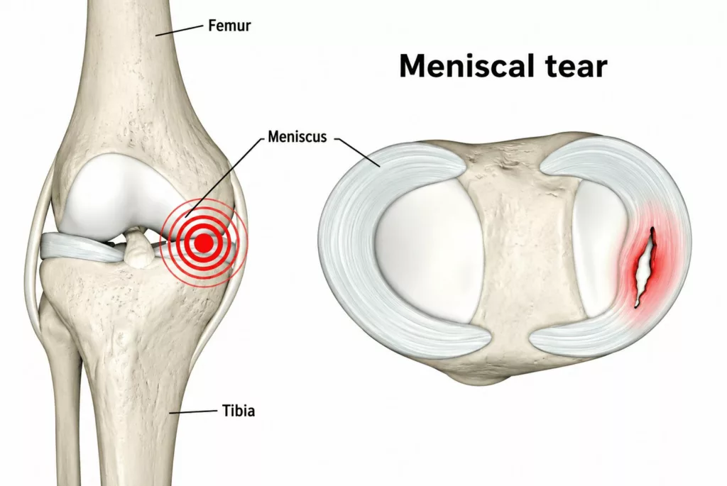
Each knee contains two distinct crescent-shaped fibrocartilaginous structures known as the medial (inner) meniscus and the lateral (outer) meniscus. Their unique anatomical configuration and resilient fibrocartilaginous composition enable them to act as essential shock absorbers whilst contributing to joint stability and ensuring optimal distribution of load across the knee. Repetitive mechanical stress, chronic microtrauma or acute high-energy injuries may lead to damage of these meniscal structures.
Clinical assessment identifies several patterns of meniscal injury, including complex fissures, tears and peripheral detachments.
Specific lesion: In certain clinical situations, a longitudinal tear extending from the posterior to the anterior horn may result in displacement of the torn fragment into the intercondylar notch. This displacement produces a mechanical blockage of the knee and is referred to as a bucket-handle tear.
Causes of Meniscal Tears
Knee Surgery Specialist | Hauts-de-Seine (92)
Meniscal lesions generally arise from two principal mechanisms.
Traumatic lesions
These predominantly affect younger, active and sporting individuals. The meniscus may sustain significant structural damage following a violent impact or a twisting injury to the knee. Such injuries commonly occur during pivoting sports involving rotational forces and direct contact, including football, alpine skiing, rugby and martial arts.
Degenerative lesions
With advancing age, the meniscal tissue undergoes progressive physiological wear and structural degeneration as a result of cumulative microtrauma over time. These degenerative changes frequently occur alongside osteoarthritis and are commonly referred to as meniscosis.
Meniscal Surgery in Rueil-Malmaison
Knee Surgery in the Hauts-de-Seine
Operation d'une chirurgie méniscale
The choice of surgical technique depends primarily on the healing potential of the meniscal lesion.
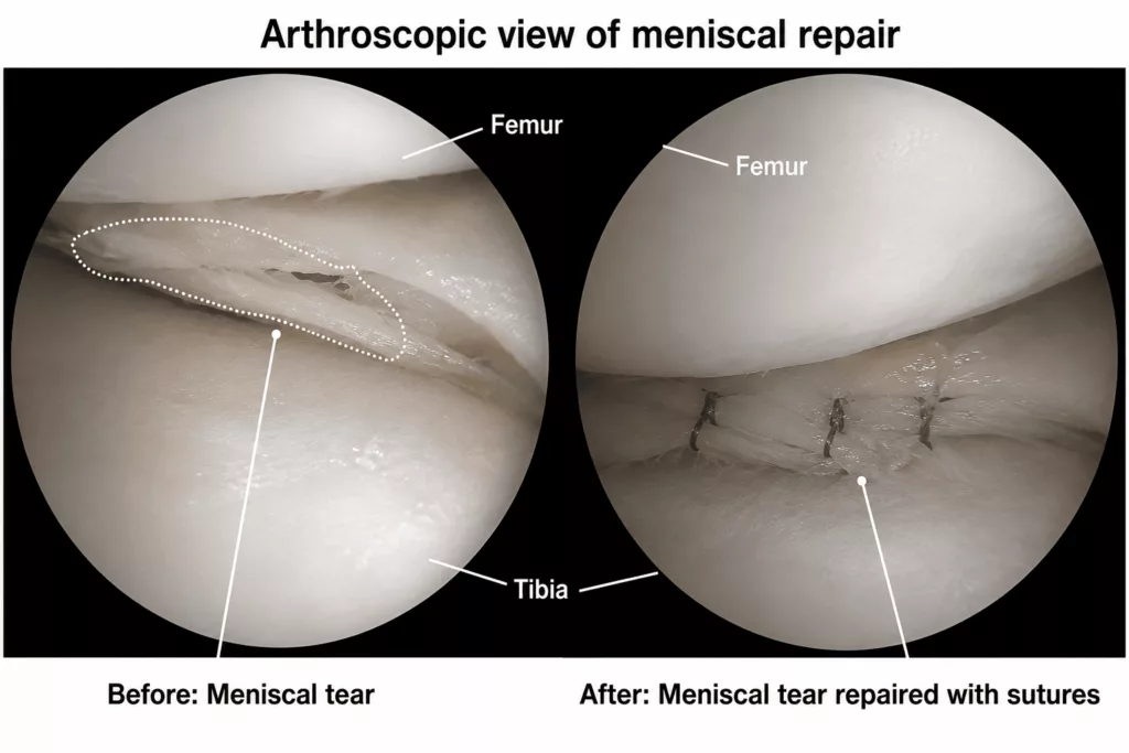
If the tear demonstrates good biological healing potential, as is typically the case in younger patients presenting with a recent traumatic tear located within the vascularised peripheral red-red zone, every effort is made to preserve the meniscus by performing a meniscal repair. The torn meniscus is repaired using high-strength orthopaedic sutures securely anchored to the joint capsule. The objective is to bring the edges of the tear into close apposition to promote biological healing and fibrocartilaginous union.
Conversely, if the lesion has no realistic healing potential, as is generally the case in older patients, chronic tears or lesions confined to the avascular white-white zone, the damaged, non-viable portion of the meniscus must be removed. This procedure is known as a meniscectomy. Whenever possible, only the damaged tissue is excised (partial meniscectomy) in order to preserve the maximum amount of healthy, functional meniscal tissue.
All procedures are performed arthroscopically. Arthroscopy is a modern minimally invasive, video-assisted surgical technique that avoids the need for an open arthrotomy. Two small 5 mm incisions are made at the front of the knee to introduce a high-definition arthroscope together with the specialised instruments required to repair or remove the damaged tissue. Compared with conventional open surgery, this technique offers several recognised advantages, including reduced post-operative pain, smaller and more discreet scars, and a faster functional recovery.
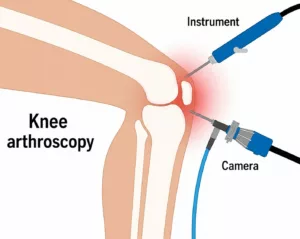
The procedure generally lasts around 30 minutes. It is performed under either general anaesthesia or spinal anaesthesia, during which only the lower limbs are anaesthetised. The choice of anaesthetic is determined jointly with the patient during the pre-operative consultation with the anaesthetist.
The operation is routinely carried out as a day-case procedure, allowing patients to return home on the day of surgery.
Clinical Indications: When is Meniscal Surgery Recommended?
In the vast majority of cases, a meniscal lesion is not considered a surgical emergency. Following the acute, highly painful phase immediately after the initial injury, conservative first-line treatment is initiated, including rest, cryotherapy (ice application), appropriate analgesia and non-steroidal anti-inflammatory drugs (NSAIDs). Once the joint swelling has subsided, patients are encouraged to resume their normal daily activities progressively over several weeks. The knee is then reassessed clinically, and if pain, mechanical symptoms or functional impairment persist, surgical treatment may be recommended.
In rare circumstances, urgent surgery is required. This occurs when the knee becomes mechanically locked, preventing full extension of the leg. This presentation is characteristic of a displaced bucket-handle tear, in which the torn meniscal fragment becomes trapped within the joint.
Why Perform Meniscal Surgery?
The natural progression of a meniscal tear is often unfavourable because the inner portion of the meniscus has a very limited blood supply, significantly reducing its capacity for spontaneous healing. When persistent pain or functional limitation remains despite conservative treatment, surgery may be indicated.
Depending on the type and location of the lesion, two surgical options are available: preservation of the meniscus by meniscal repair, or selective removal of the damaged tissue by partial meniscectomy. In both cases, the primary objective is to relieve pain, eliminate mechanical locking and allow the patient to return to normal daily activities and sporting activities.
Post-operative Care and Rehabilitation
As this procedure is performed as a day-case operation, patients return home on the day of surgery.
The dressings should be changed every two days for approximately two weeks. A personalised multimodal pain management programme is prescribed to ensure optimal comfort and facilitate early mobilisation. Oral anticoagulant treatment is routinely prescribed for two weeks to reduce the risk of deep vein thrombosis (DVT). This prophylactic treatment does not require routine blood monitoring.
The rehabilitation programme depends on the procedure performed.
Following meniscal repair, patients should use two crutches with partial weight-bearing for approximately four weeks. This protected weight-bearing is essential to allow the repaired meniscus to heal successfully. Physiotherapy should begin immediately and continue for approximately two months, focusing on maintaining quadriceps strength, preserving knee mobility and restoring proprioception. Return to work and driving is generally possible after around one month, depending on occupational demands. Sporting activities should not be resumed before approximately three months to allow complete biological healing.
Following a partial meniscectomy, rehabilitation is considerably less restrictive. Full weight-bearing is permitted immediately as tolerated, without walking aids in most cases. Physiotherapy should also begin immediately and continue for approximately two months to restore muscle strength and normal range of movement. Driving and return to work are usually possible after around two weeks, whilst sporting activities may gradually resume after approximately two months.
These recovery times are indicative only and should always be adapted according to the individual’s rate of healing, tissue quality and functional progress.
Long-term clinical and radiographic follow-up is recommended to monitor knee function and identify any early signs of degenerative joint changes.
Risks and Potential Complications
Although every precaution is taken to ensure the safest possible outcome, all surgical procedures carry a risk of complications. Whilst uncommon, the following complications may occur:
Deep Vein Thrombosis (DVT) and Pulmonary Embolism
A rare thromboembolic complication, the risk of which is reduced by routine anticoagulant treatment during the first two weeks following surgery.
Joint Infection (Septic Arthritis)
An uncommon complication, occurring in fewer than 1% of cases. If infection develops, urgent arthroscopic joint washout combined with prolonged targeted antibiotic therapy is required.
Arthrofibrosis and Joint Stiffness
Loss of knee movement may occur, particularly if the post-operative physiotherapy programme is not followed appropriately.
Haematoma / Haemarthrosis
Bleeding within the joint may lead to a painful haemarthrosis. Depending on its severity, treatment may involve aspiration, surgical drainage or, exceptionally, blood transfusion.
Nerve Injury
Small superficial sensory nerves, particularly the infrapatellar branch of the saphenous nerve, may occasionally be injured. This may result in numbness or altered sensation around the surgical scars.
Failure of Meniscal Healing
Despite technically successful meniscal repair, biological healing does not occur in every case. Failure of healing may lead to recurrent pain or mechanical symptoms, making further arthroscopic surgery necessary, either to repeat the repair or to perform a partial meniscectomy.
Complex Regional Pain Syndrome (CRPS)
Complex Regional Pain Syndrome is a rare neurovascular condition characterised by persistent pain that is disproportionate to the initial injury, together with disturbances in circulation and nerve function. Management may require specialist multidisciplinary care, prolonged physiotherapy and pain management. The onset, duration and long-term outcome remain unpredictable.
This list summarises the principal complications associated with meniscal surgery but is not exhaustive. Other rare complications may occur. Your surgeon will discuss the risks relevant to your individual circumstances during your consultation.
Results of Meniscal Surgery
Following either meniscal repair or partial meniscectomy, pain and mechanical symptoms usually improve rapidly after surgery. Recovery of muscle strength and restoration of full knee mobility generally occur within two to three months.
It is important to recognise that partial meniscectomy permanently removes part of the meniscus, which functions as a shock absorber within the knee. This inevitably alters joint biomechanics and increases the long-term risk of developing knee osteoarthritis, with the degree of risk depending on the amount and location of meniscal tissue removed.
Following meniscal repair, successful healing cannot be guaranteed in every patient. Published clinical studies report healing rates of approximately 60% for the medial meniscus and up to 75% for the lateral meniscus. If healing fails, recurrent pain or mechanical symptoms may develop, requiring further arthroscopic surgery to remove the non-healed portion of the meniscus.