Meniscal Tear in Hauts-de-Seine
Knee Conditions
What Is a Meniscal Tear?
Dr Simon Tournemine in Hauts-de-Seine (92)
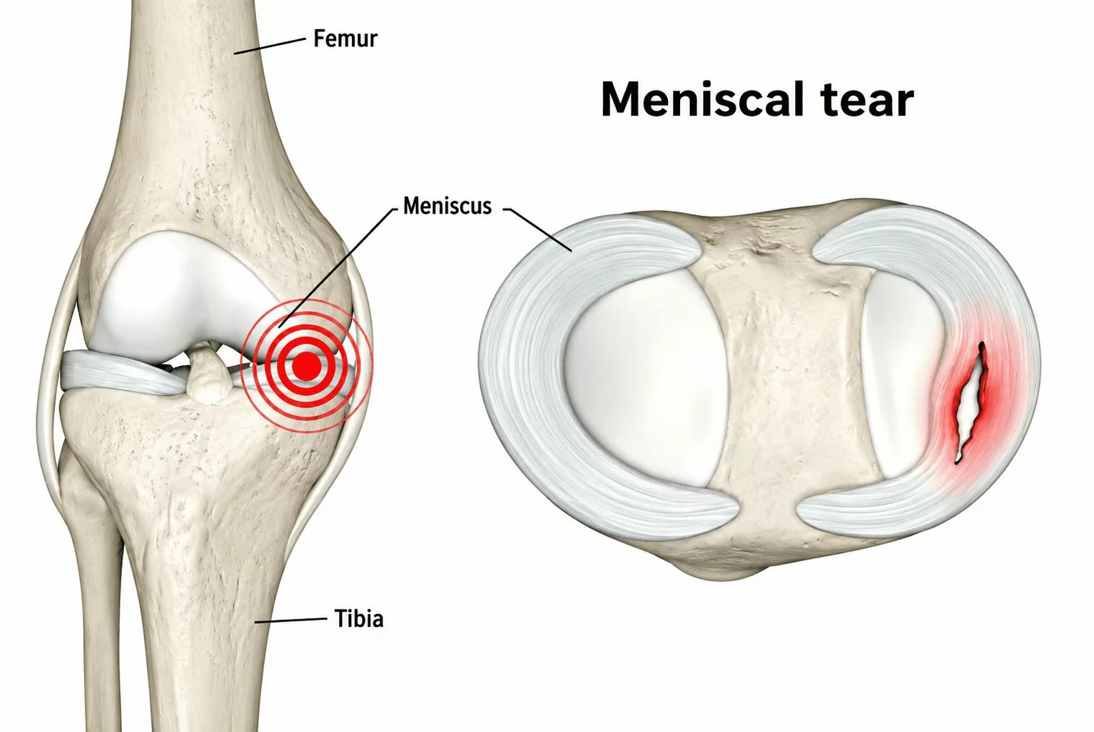
Each knee contains two menisci: the medial (inner) meniscus and the lateral (outer) meniscus. These crescent-shaped fibrocartilaginous structures act as natural shock absorbers within the knee joint while also contributing to joint stability, load distribution and smooth movement.
Repeated mechanical stress or a sudden traumatic injury can damage the menisci, resulting in different types of lesions, including:
- Meniscal fissures
- Meniscal tears
- Meniscal root or peripheral detachments
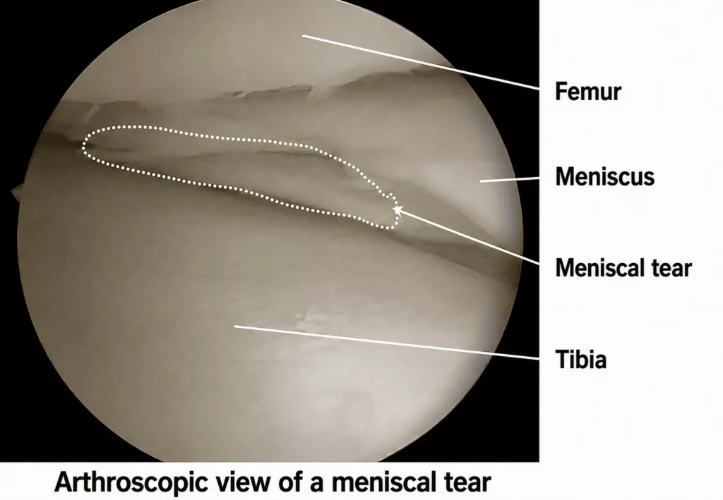
In some cases, a tear completely divides the meniscus from front to back. The detached fragment may then displace into the centre of the knee joint and become trapped within the intercondylar notch. This type of injury is known as a bucket-handle tear.
Causes and Risk Factors for a Meniscal Tear
Knee Specialist in Hauts-de-Seine (92)
There are two main categories of meniscal injury: traumatic tears and degenerative tears.
Traumatic meniscal tears occur predominantly in young and physically active individuals. They usually result from abnormal twisting movements or direct trauma to the knee, particularly during so-called pivot-contact sports, such as football, skiing, rugby and combat sports. The most common mechanisms of injury include twisting the knee while the foot remains firmly planted on the ground or suddenly rising from a deep squatting position.
Degenerative meniscal tears develop progressively as the meniscus undergoes age-related wear. Throughout life, repeated microtrauma gradually weakens the fibrocartilage, making it more susceptible to tearing. These degenerative changes, often referred to as meniscosis, commonly occur alongside knee osteoarthritis and are more frequently observed in former athletes and individuals whose occupations involve heavy physical work.
Meniscal Tear in Rueil-Malmaison
Knee Surgery in Hauts-de-Seine
Symptoms of a Meniscal Tear
In younger patients with a traumatic meniscal injury, pain usually occurs suddenly following a twisting movement of the knee or when rising abruptly from a squatting position. Sporting activity generally has to be stopped immediately, and the knee may become swollen because bleeding occurs within the joint (haemarthrosis). In some cases, a displaced bucket-handle tear may cause the knee to lock, making it impossible to fully straighten the leg.
In older patients, where the meniscus and the articular cartilage have gradually worn over time, pain typically develops during physical activity or when pivoting on the affected leg. A limp may gradually appear, occasionally making the use of a walking stick necessary. Symptoms usually progress slowly, with painful flare-ups alternating with periods of relative improvement.
Without appropriate treatment, symptoms often persist and vary according to the severity and extent of the meniscal injury. They may include:
- Persistent knee pain
- Recurrent episodes of knee swelling
- Clicking or cracking sensations within the knee
- Knee locking with an inability to fully extend the joint
Preventing a Meniscal Tear
Maintaining good knee flexibility together with adequate muscle strength helps reduce the risk of meniscal injuries. A thorough warm-up before sporting activity, combined with regular muscle-strengthening and stretching exercises, plays an important role in protecting the knee.
Diagnosis of a Meniscal Tear
The consultation begins with a detailed medical history to establish the mechanism of injury and evaluate the patient’s symptoms, including pain, clicking sensations, locking episodes and feelings of instability. The type of sport practised and the patient’s level of physical activity are also taken into consideration.
Clinical examination includes specific manoeuvres designed to compress the meniscus in order to reproduce pain and identify the injured area. Knee mobility and overall stability are also carefully assessed by examining the anterior and posterior cruciate ligaments together with the medial and lateral collateral ligaments.
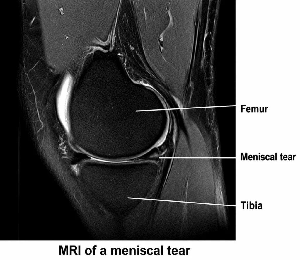
Imaging studies are routinely performed alongside the clinical examination. Standard knee X-rays help identify associated conditions such as fractures or osteoarthritis. Magnetic resonance imaging (MRI) is the reference investigation, allowing precise assessment of the meniscus, the location and pattern of the tear, and the presence of associated ligament injuries, which are commonly seen in conjunction with meniscal lesions.
Treatment of a Meniscal Tear
The natural progression of a meniscal tear is generally unfavourable because the meniscus has a limited blood supply and therefore heals poorly without treatment. When symptoms such as persistent pain, swelling or mechanical locking continue despite conservative management, surgical treatment may be recommended.
The choice of procedure depends on the type, location and extent of the tear. Whenever possible, the meniscus is preserved by performing a meniscal repair. When repair is not feasible, only the damaged portion of the meniscus is removed (partial meniscectomy) while preserving as much healthy tissue as possible.
The objective of surgery is to relieve pain, eliminate locking and recurrent swelling, restore normal knee function and allow a safe return to everyday activities and sporting practice.