
Patellar Tendinopathy in Hauts-de-Seine
Knee Surgery
What Is Patellar Tendinopathy?
Dr Simon Tournemine in Hauts-de-Seine (92)
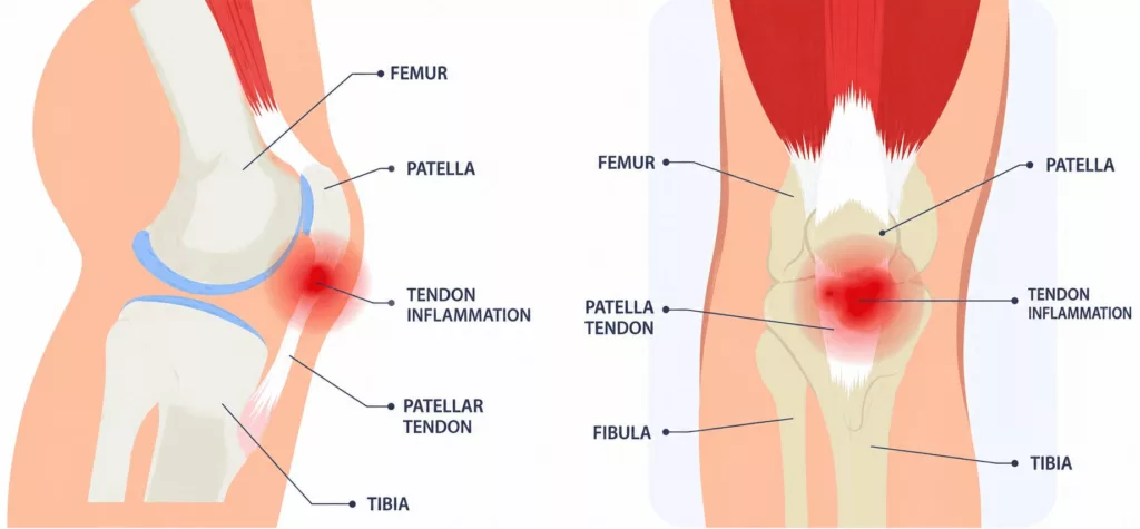
The knee joint is formed by three bones: the patella (kneecap), the femur and the tibia. Knee extension is produced by the extensor mechanism, which is powered by the quadriceps muscle. As the quadriceps contracts, it pulls on the patella, which is connected to the tibia via the patellar tendon. Because it transmits substantial mechanical forces during walking, running and jumping, the patellar tendon is subjected to considerable stress.
Patellar tendinopathy, commonly referred to as jumper’s knee, results from the accumulation of repeated microscopic injuries within the patellar tendon between the patella and the tibia. Repetitive overuse of the knee causes tiny tears to develop within the tendon fibres. As these microtears accumulate, the body attempts to repair the damaged tissue, leading to local inflammation and progressive weakening of the tendon. When this process persists over time, chronic patellar tendinopathy develops.
Causes and Risk Factors for Patellar Tendinopathy
Knee Surgery Specialist in Hauts-de-Seine (92)
Patellar tendinopathy is particularly common in young athletes participating in sports that involve frequent jumping, such as basketball, handball, football and volleyball.
Several factors may increase the risk of developing this condition, including:
- A sudden increase in training intensity or training volume
- Repeated training on hard surfaces
- Inappropriate footwear
- Poor hydration
- Tobacco and excessive alcohol consumption
- A history of Osgood-Schlatter disease during childhood
Patellar Tendinopathy in Rueil-Malmaison
Knee Surgery in Hauts-de-Seine
Symptoms of Patellar Tendinopathy
Pain is the principal symptom of patellar tendinopathy. It is typically localised to the front of the knee, between the lower pole of the patella and the tibia. The knee may also become swollen and warm due to local inflammation.
Initially, pain occurs only during sporting activity. As the condition progresses, symptoms become increasingly frequent and may eventually be present during everyday activities or even at rest. Daily tasks such as climbing stairs or rising from a chair may become difficult, while sporting performance is progressively limited.
In advanced and uncommon cases, chronic tendon degeneration may ultimately result in patellar tendon rupture.
Preventing Patellar Tendinopathy
Maintaining good knee flexibility together with adequate muscle strength helps reduce the risk of patellar tendinopathy. A thorough warm-up before exercise, combined with regular strengthening and stretching exercises, is essential.
Training intensity should always be increased progressively to avoid excessive tendon loading. Maintaining a healthy lifestyle is equally important. Adequate hydration, limiting alcohol consumption and avoiding smoking are recognised protective measures that contribute to tendon health and reduce the risk of chronic tendinopathy.
Diagnosis of Patellar Tendinopathy
The consultation begins with a detailed medical history to identify the factors responsible for the pain and to assess its impact on sporting activities and daily life.
Clinical examination focuses on identifying tenderness over the patellar tendon and detecting any associated joint effusion. Functional tests, including jumping manoeuvres and single-leg weight-bearing, are performed to reproduce the patient’s symptoms. Knee stability is also assessed to identify any associated meniscal or ligament injuries.
Standard knee X-rays may be requested to identify calcification or ossification within the patellar tendon.
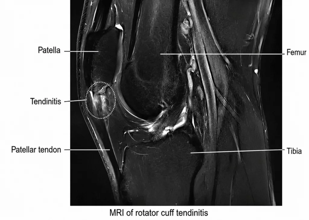
Magnetic resonance imaging (MRI) remains the reference investigation, providing direct visualisation of the tendon lesion, its extent and severity. MRI also enables the identification of any associated abnormalities affecting the knee.
Treatment of Patellar Tendinopathy
Initial treatment is conservative and focuses on reducing pain while promoting tendon healing. Management includes temporary reduction or modification of sporting activities together with anti-inflammatory medication when appropriate.
Contributing factors should also be addressed by optimising footwear, maintaining adequate hydration, limiting alcohol consumption and smoking, and following a structured rehabilitation programme based on progressive stretching and strengthening exercises before returning to sport.
Corticosteroid injections are generally avoided, as they may weaken the tendon and increase the risk of tendon rupture.
When conservative treatment fails to provide sufficient improvement, surgical management may be considered. The recommended procedure is patellar tendon debridement (patellar tendon scraping).