
Tarsal Tunnel Release in Hauts-de-Seine

Foot Surgery

What is a tarsal tunnel release?

Dr Simon Tournemine in Hauts-de-Seine (92)

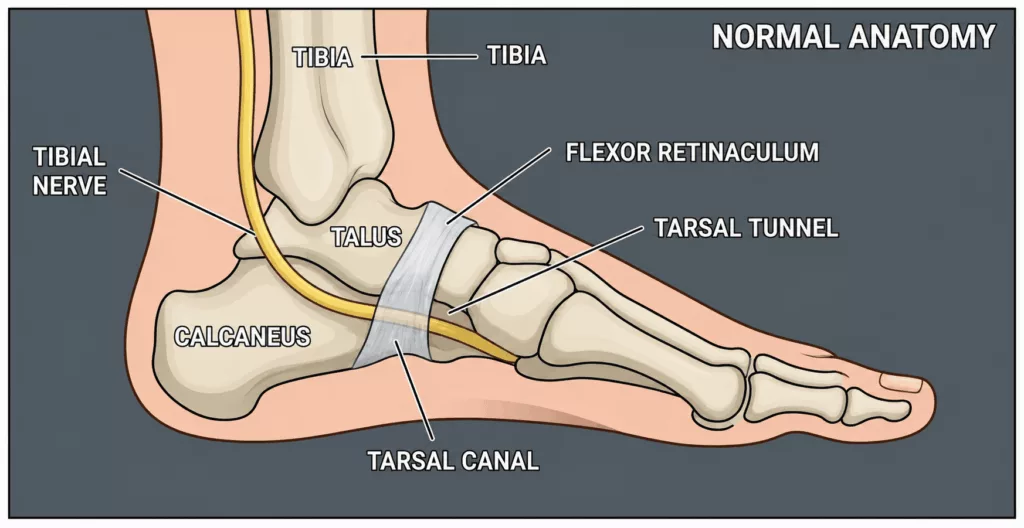
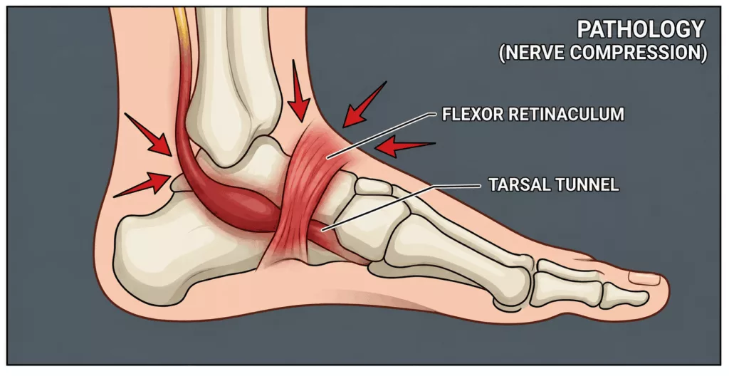
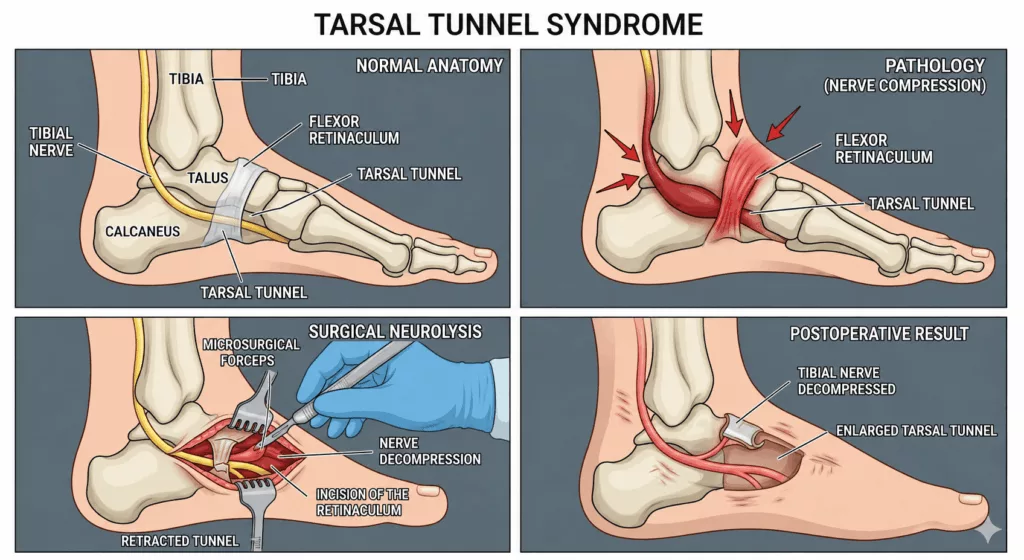
Tarsal tunnel syndrome is caused by compression of the posterior tibial nerve, or one of its terminal branches, as it passes through a narrow osteofibrous tunnel located on the inner side of the ankle, beneath the medial malleolus.

This nerve compression causes disabling neuropathic pain, burning sensations, tingling, numbness and electric shock-like sensations affecting the sole of the foot, the heel and the toes. Symptoms are typically aggravated by walking, prolonged standing and frequently worsen at night.

Tarsal tunnel release (neurolysis) is the surgical procedure performed to decompress the nerve by dividing the flexor retinaculum (laciniate ligament), thereby relieving pressure on the nerve and restoring optimal nerve conduction.

Tarsal Tunnel Release

Specialist in Foot Surgery in Hauts-de-Seine (92)

The objective of surgery is to permanently decompress the posterior tibial nerve in order to relieve pain, restore nerve function and prevent irreversible nerve damage.

Several surgical techniques may be considered depending on the underlying cause and individual anatomy.

Tarsal Tunnel Release in Rueil-Malmaison

Foot Surgery in Hauts-de-Seine

Conventional open or minimally invasive approach

A curved vertical incision measuring approximately 4 to 5 cm is made behind and below the medial malleolus.

The surgeon divides the flexor retinaculum, which forms the roof of the tarsal tunnel, allowing complete decompression of the posterior tibial nerve as far as its division into the medial and lateral plantar nerves.

If a local compressive lesion is identified, such as a ganglion cyst, enlarged veins or an accessory muscle, it is removed during the same procedure.

Endoscopic technique

In selected cases, decompression may be performed using an endoscopic technique through very small incisions under video guidance.

The most appropriate surgical approach depends on the anatomy of the ankle and the cause of the nerve compression and will be discussed during your consultation.

The operation generally lasts 20 to 30 minutes.

It is performed under either general anaesthesia or regional anaesthesia, where only the lower limb is numbed. The choice of anaesthesia is made together with the anaesthetist during your preoperative assessment.

The procedure is carried out as day-case (outpatient) surgery, allowing you to return home on the same day.

Except in exceptional circumstances, both feet are not usually operated on simultaneously, in order to preserve mobility during recovery.
Conservative treatment is always attempted before surgery.

This usually includes:

  • rest and activity modification;
  • custom-made foot orthoses to correct underlying biomechanical abnormalities such as pes planovalgus;
  • ultrasound-guided corticosteroid injections;
  • physiotherapy.

If symptoms persist despite several months of appropriate conservative management, or if electromyography (EMG) demonstrates objective evidence of significant nerve compression, surgical decompression is recommended.

Surgery is never performed as a preventive procedure in the absence of disabling symptoms.

The operation is performed as day-case surgery, allowing discharge home on the day of the procedure.

A sterile dressing is applied in theatre and should be inspected and renewed every two days until skin healing is complete, usually after approximately two weeks.

Pain management is carefully supervised to maximise comfort and facilitate early mobilisation.

Walking is generally permitted within the first few days using comfortable footwear, such as trainers, with temporary use of crutches if required for pain relief.

Following surgery, swelling of the foot and ankle is common and expected. Rest, ice application and elevation of the limb help reduce postoperative oedema.

A programme of physiotherapy is prescribed to restore ankle mobility, mobilise the scar to minimise nerve adhesions and reduce swelling.

Time away from work is generally three to four weeks, although this varies according to occupation. Patients with predominantly office-based work may return sooner.

High-impact sporting activities are usually resumed between two and three months after surgery.

These recovery times are average estimates and vary according to individual healing.

Regular postoperative clinical follow-up is essential to monitor recovery of sensation and foot function.

Every precaution is taken to promote optimal recovery. Nevertheless, as with any surgical procedure, complications may occur, although they remain uncommon.

Potential risks include:

  • deep vein thrombosis (DVT) and pulmonary embolism, minimised through early mobilisation or anticoagulant treatment where indicated according to individual risk factors;
  • worsening of pre-existing medical conditions, including diabetes or cardiovascular disease, monitored by the anaesthetic team;
  • infection (risk below 1%), occasionally requiring further surgery together with antibiotic treatment;
  • haematoma, with strict adherence to postoperative elevation recommendations helping to reduce this risk;
  • persistent nerve irritation or delayed neurological recovery. Because the nerve has often been compressed for a prolonged period before surgery, pain, paraesthesia or numbness may require several months to improve and, in some cases, may not completely resolve. Rarely, adjacent sensory nerve branches may be stretched or injured during the procedure;
  • delayed wound healing or scar adhesions. The skin on the inner aspect of the ankle is relatively thin, and scar tissue may occasionally tether the nerve, requiring dedicated scar mobilisation therapy;
  • Complex Regional Pain Syndrome (CRPS), a rare inflammatory pain syndrome that remains poorly understood. It is treated medically and may persist for several months, or occasionally several years, requiring specialised rehabilitation, further investigations and, in some cases, management by a dedicated pain clinic. Its occurrence and progression remain unpredictable.

These represent the principal potential complications, although this list is not exhaustive. Your surgeon will discuss your individual circumstances during your consultation.

Without treatment, tarsal tunnel syndrome generally progresses, potentially resulting in persistent pain, permanent loss of sensation affecting the sole of the foot and wasting of the intrinsic foot muscles.

The objectives of surgery are twofold:

  • to relieve neuropathic pain, including burning sensations and electric shock-like symptoms, both at rest and during walking;
  • to prevent further nerve deterioration and preserve long-term sensory and motor function of the foot.

Most patients experience a significant reduction in pain following surgery. However, the speed and extent of neurological recovery depend largely on the duration and severity of nerve compression before the operation, as nerve regeneration is inherently a slow biological process.